7CNZ

Crystal structure of 10PE bound PSD from E. coli (2.70 A)


Experimental Data Snapshot

  • Method: X-RAY DIFFRACTION
  • Resolution: 2.70 Å
  • R-Value Free: 0.276 
  • R-Value Work: 0.227 
  • R-Value Observed: 0.230 

wwPDB Validation   3D Report Full Report


Ligand Structure Quality Assessment 


This is version 2.0 of the entry. See complete history


Literature

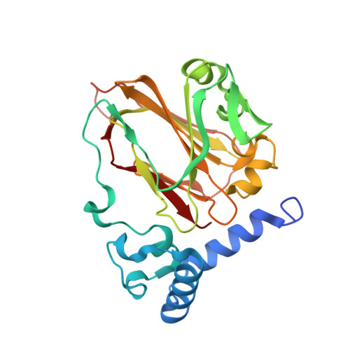
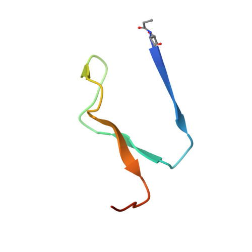
Structural insights into phosphatidylethanolamine formation in bacterial membrane biogenesis.

Cho, G.Lee, E.Kim, J.

(2021) Sci Rep 11: 5785-5785

  • DOI: https://doi.org/10.1038/s41598-021-85195-5
  • Primary Citation of Related Structures:  
    7CNW, 7CNX, 7CNY, 7CNZ

  • PubMed Abstract: 

    Phosphatidylethanolamine (PE), a major component of the cellular membrane across all domains of life, is synthesized exclusively by membrane-anchored phosphatidylserine decarboxylase (PSD) in most bacteria. The enzyme undergoes auto-cleavage for activation and utilizes the pyruvoyl moiety to form a Schiff base intermediate with PS to facilitate decarboxylation. However, the structural basis for self-maturation, PS binding, and decarboxylation processes directed by PSD remain unclear. Here, we present X-ray crystal structures of PSD from Escherichia coli, representing an apo form and a PE-bound complex, in which the phospholipid is chemically conjugated to the essential pyruvoyl residue, mimicking the Schiff base intermediate. The high-resolution structures of PE-complexed PSD clearly illustrate extensive hydrophobic interactions with the fatty acyl chains of the phospholipid, providing insights into the broad specificity of the enzyme over a wide range of cellular PS. Furthermore, these structures strongly advocate the unique topology of the enzyme in a lipid bilayer environment, where the enzyme associates with cell membranes in a monotopic fashion via the N-terminal domain composed of three amphipathic helices. Lastly, mutagenesis analyses reveal that E. coli PSD primarily employs D90/D142-H144-S254 to achieve auto-cleavage for the proenzyme maturation, where D90 and D142 act in complementary to each other.


  • Organizational Affiliation

    Department of Chemistry, Gwangju Institute of Science and Technology, Gwangju, 61005, Republic of Korea.


Macromolecules
Find similar proteins by:  (by identity cutoff)  |  3D Structure
Entity ID: 1
MoleculeChains Sequence LengthOrganismDetailsImage
Phosphatidylserine decarboxylase beta chain
A, C, E, G
253Escherichia coli K-12Mutation(s): 0 
Gene Names: psdFAZ83_09855
EC: 4.1.1.65
Membrane Entity: Yes 
UniProt
Find proteins for P0A8K1 (Escherichia coli (strain K12))
Explore P0A8K1 
Go to UniProtKB:  P0A8K1
Entity Groups  
Sequence Clusters30% Identity50% Identity70% Identity90% Identity95% Identity100% Identity
UniProt GroupP0A8K1
Sequence Annotations
Expand
  • Reference Sequence
Find similar proteins by:  (by identity cutoff)  |  3D Structure
Entity ID: 2
MoleculeChains Sequence LengthOrganismDetailsImage
Phosphatidylserine decarboxylase alpha chain
B, D, F, H
42Escherichia coli K-12Mutation(s): 1 
Gene Names: psdFAZ83_09855
EC: 4.1.1.65
Membrane Entity: Yes 
UniProt
Find proteins for P0A8K1 (Escherichia coli (strain K12))
Explore P0A8K1 
Go to UniProtKB:  P0A8K1
Entity Groups  
Sequence Clusters30% Identity50% Identity70% Identity90% Identity95% Identity100% Identity
UniProt GroupP0A8K1
Sequence Annotations
Expand
  • Reference Sequence
Experimental Data & Validation

Experimental Data

  • Method: X-RAY DIFFRACTION
  • Resolution: 2.70 Å
  • R-Value Free: 0.276 
  • R-Value Work: 0.227 
  • R-Value Observed: 0.230 
  • Space Group: P 21 21 21
Unit Cell:
Length ( Å )Angle ( ˚ )
a = 79.723α = 90
b = 102.373β = 90
c = 168.993γ = 90
Software Package:
Software NamePurpose
REFMACrefinement
MxDCdata collection
XDSdata reduction
Aimlessdata scaling
MOLREPphasing
Cootmodel building
PDB_EXTRACTdata extraction

Structure Validation

View Full Validation Report


Ligand Structure Quality Assessment 


Entry History & Funding Information

Deposition Data


Funding OrganizationLocationGrant Number
National Research Foundation (NRF, Korea)Korea, Republic Of--

Revision History  (Full details and data files)

  • Version 1.0: 2021-03-24
    Type: Initial release
  • Version 2.0: 2023-11-15
    Changes: Atomic model, Data collection, Database references, Derived calculations, Refinement description