DeSiphering receptor core-induced and ligand-dependent conformational changes in arrestin via genetic encoded trimethylsilyl 1 H-NMR probe.
Liu, Q., He, Q.T., Lyu, X., Yang, F., Zhu, Z.L., Xiao, P., Yang, Z., Zhang, F., Yang, Z.Y., Wang, X.Y., Sun, P., Wang, Q.W., Qu, C.X., Gong, Z., Lin, J.Y., Xu, Z., Song, S.L., Huang, S.M., Guo, S.C., Han, M.J., Zhu, K.K., Chen, X., Kahsai, A.W., Xiao, K.H., Kong, W., Li, F.H., Ruan, K., Li, Z.J., Yu, X., Niu, X.G., Jin, C.W., Wang, J., Sun, J.P.(2020) Nat Commun 11: 4857-4857
- PubMed: 32978402 Search on PubMedSearch on PubMed Central
- DOI: https://doi.org/10.1038/s41467-020-18433-5
- Primary Citation Related Structures:
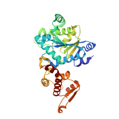
6KRG, 7CKG, 7CKH - PubMed Abstract:
Characterization of the dynamic conformational changes in membrane protein signaling complexes by nuclear magnetic resonance (NMR) spectroscopy remains challenging. Here we report the site-specific incorporation of 4-trimethylsilyl phenylalanine (TMSiPhe) into proteins, through genetic code expansion. Crystallographic analysis revealed structural changes that reshaped the TMSiPhe-specific amino-acyl tRNA synthetase active site to selectively accommodate the trimethylsilyl (TMSi) group. The unique up-field 1 H-NMR chemical shift and the highly efficient incorporation of TMSiPhe enabled the characterization of multiple conformational states of a phospho-β2 adrenergic receptor/β-arrestin-1(β-arr1) membrane protein signaling complex, using only 5 μM protein and 20 min of spectrum accumulation time. We further showed that extracellular ligands induced conformational changes located in the polar core or ERK interaction site of β-arr1 via direct receptor transmembrane core interactions. These observations provided direct delineation and key mechanism insights that multiple receptor ligands were able to induce distinct functionally relevant conformational changes of arrestin.
- Institute of Biophysics, Chinese Academy of Sciences, 15 Datun Road, Chaoyang district, Beijing, 100101, China.
Organizational Affiliation: