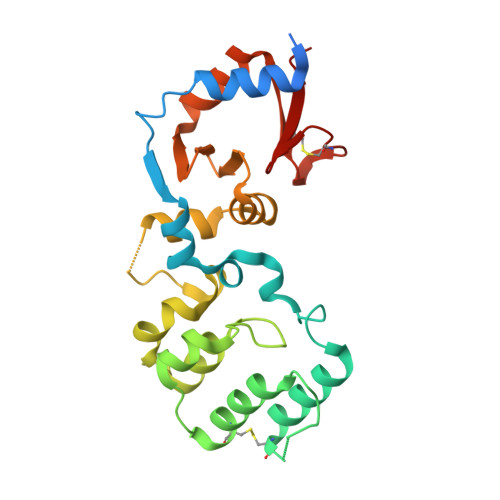
Crystal structure of the periplasmic sensor domain of histidine kinase VbrK suggests indirect sensing of beta-lactam antibiotics.
Goh, B.C., Chua, Y.K., Qian, X., Lin, J., Savko, M., Dedon, P.C., Lescar, J.(2020) J Struct Biol 212: 107610-107610
- PubMed: 32890780 Search on PubMed
- DOI: https://doi.org/10.1016/j.jsb.2020.107610
- Primary Citation Related Structures:
7CJR - PubMed Abstract:
Bacterial two-component regulatory systems (TCS) play important roles in sensing environmental stimuli and responding to them by regulating gene expression. VbrK/VbrR, a TCS in Vibrio parahaemolyticus, confers resistance to β-lactam antibiotics through activating a β-lactamase gene. Its periplasmic sensor domain was previously suggested to detect β-lactam antibiotics by direct binding. Here, we report a crystal structure of the periplasmic sensing domain of VbrK (VbrK SD ) using sulfur-based single-wavelength anomalous diffraction (S-SAD) phasing. Contrary to most bacterial sensor domains which form dimers, we show that VbrK SD is a monomer using size exclusion chromatography coupled with multi-angle light scattering. This observation is also supported by molecular dynamics simulations. To quantify the binding affinity of β-lactam antibiotics to VbrK SD , we performed isothermal titration calorimetry and other biophysical analyses. Unexpectedly, VbrK SD did not show any significant binding to β-lactam antibiotics. Therefore, we propose that the detection of β-lactam antibiotics by VbrK is likely to be indirect via an as yet unidentified mechanism.
- Antimicrobial Resistance Interdisciplinary Research Group, Singapore-MIT Alliance for Research and Technology Centre, 138602, Singapore; NTU Institute of Structural Biology, Nanyang Technological University, 636921, Singapore. Electronic address: boonchong@smart.mit.edu.
Organizational Affiliation: