Atomic differentiation of silver binding preference in protein targets: Escherichia coli malate dehydrogenase as a paradigm.
Wang, H., Yang, X., Wang, M., Hu, M., Xu, X., Yan, A., Hao, Q., Li, H., Sun, H.(2020) Chem Sci 11: 11714-11719
- PubMed: 34123202 Search on PubMedSearch on PubMed Central
- DOI: https://doi.org/10.1039/d0sc04151c
- Primary Citation Related Structures:
7CGC, 7CGD - PubMed Abstract:
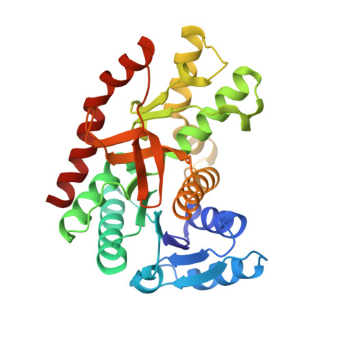
Understanding how metallodrugs interact with their protein targets is of vital importance for uncovering their molecular mode of actions as well as overall pharmacological/toxicological profiles, which in turn facilitates the development of novel metallodrugs. Silver has been used as an antimicrobial agent since antiquity, yet there is limited knowledge about silver-binding proteins. Given the multiple dispersed cysteine residues and histidine-methionine pairs, Escherichia coli malate dehydrogenase ( Ec MDH) represents an excellent model to investigate silver coordination chemistry as well as its targeting sites in enzymes. We show by systematic biochemical characterizations that silver ions (Ag + ) bind Ec MDH at multiple sites including three cysteine-containing sites. By X-ray crystallography, we unravel the binding preference of Ag + to multiple binding sites in Ec MDH, i.e., Cys113 > Cys251 > Cys109 > Met227. Silver exhibits preferences to the donor atoms and residues in the order of S > N > O and Cys > Met > His > Lys > Val, respectively, in Ec MDH. For the first time, we report the coordination of silver to a lysine in proteins. Besides, we also observed argentophilic interactions (Ag⋯Ag, 2.7 to 3.3 Å) between two silver ions coordinating to one thiolate. Combined with site-directed mutagenesis and an enzymatic activity test, we unveil that the binding of Ag + to the site IV (His177-Ag-Met227 site) plays a vital role in Ag + -mediated MDH inactivation. This work stands as the first unusual and explicit study of silver binding preference to multiple binding sites in its authentic protein target at the atomic resolution. These findings enrich our knowledge on the biocoordination chemistry of silver(i), which in turn facilitates the prediction of the unknown silver-binding proteins and extends the pharmaceutical potentials of metal-based drugs.
- Department of Chemistry, CAS-HKU Joint Laboratory of Metallomics on Health and Environment, The University of Hong Kong Pokfulam Road Hong Kong P. R. China haibo_wang@connect.hku.hk hsun@hku.hk.
Organizational Affiliation: