


Crystal structure of RsmG methyltransferase of M. tuberculosis
Bijpuria, S., Maurya, A., Kumar, P., Sharma, R., Taneja, B.To be published.
Experimental Data Snapshot
Entity ID: 1 | |||||
|---|---|---|---|---|---|
| Molecule | Chains | Sequence Length | Organism | Details | Image |
| Ribosomal RNA small subunit methyltransferase G | 230 | Mycobacterium tuberculosis H37Rv | Mutation(s): 0 Gene Names: rsmG, gidB, Rv3919c, MTV028.10c EC: 2.1.1 | 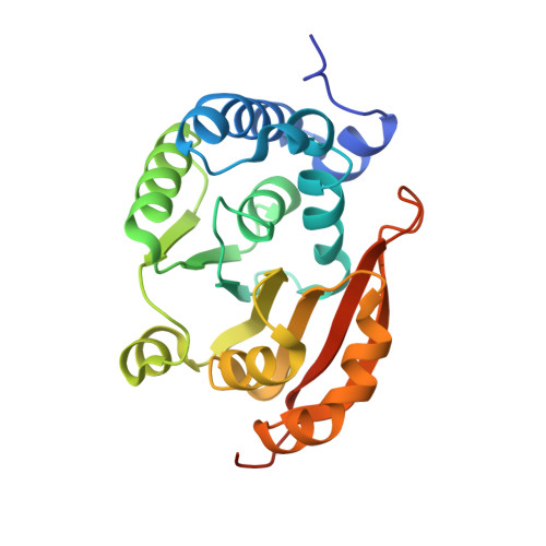 | |
UniProt | |||||
Entity Groups | |||||
| Sequence Clusters | 30% Identity50% Identity70% Identity90% Identity95% Identity100% Identity | ||||
| UniProt Group | P9WGW9 | ||||
Sequence AnnotationsExpand | |||||
Reference Sequence | |||||
| Ligands 3 Unique | |||||
|---|---|---|---|---|---|
| ID | Chains | Name / Formula / InChI Key | 2D Diagram | 3D Interactions | |
| SFG (Subject of Investigation/LOI) Download:Ideal Coordinates CCD File | B [auth A] | SINEFUNGIN C15 H23 N7 O5 LMXOHSDXUQEUSF-YECHIGJVSA-N |  | ||
| PO4 Download:Ideal Coordinates CCD File | C [auth A] | PHOSPHATE ION O4 P NBIIXXVUZAFLBC-UHFFFAOYSA-K |  | ||
| IPA Download:Ideal Coordinates CCD File | D [auth A] | ISOPROPYL ALCOHOL C3 H8 O KFZMGEQAYNKOFK-UHFFFAOYSA-N |  | ||
| Length ( Å ) | Angle ( ˚ ) |
|---|---|
| a = 44.98 | α = 90 |
| b = 63.88 | β = 90 |
| c = 72.39 | γ = 90 |
| Software Name | Purpose |
|---|---|
| PHENIX | refinement |
| iMOSFLM | data reduction |
| iMOSFLM | data scaling |
| AutoSol | phasing |
| Funding Organization | Location | Grant Number |
|---|---|---|
| Department of Science & Technology (DST, India) | India | EMR/2016/003589 |


