7CF6 | pdb_00007cf6

Crystal structure of Beta-aspartyl dipeptidase from thermophilic keratin degrading Fervidobacterium islandicum AW-1 in complex with beta-Asp-Leu dipeptide


Experimental Data Snapshot

  • Method: X-RAY DIFFRACTION
  • Resolution: 2.75 Å
  • R-Value Free: 
    0.278 (Depositor), 0.275 (DCC) 
  • R-Value Work: 
    0.196 (Depositor), 0.197 (DCC) 
  • R-Value Observed: 
    0.200 (Depositor) 

Starting Model: experimental
View more details

wwPDB Validation 3D Report Full Report

Validation slider image for 7CF6

Ligand Structure Quality Assessment 


This is version 1.2 of the entry. See complete history

Literature

Functional Characterization of Primordial Protein Repair Enzyme M38 Metallo-Peptidase From Fervidobacterium islandicum AW-1.

La, J.W.Dhanasingh, I.Jang, H.Lee, S.H.Lee, D.W.

(2020) Front Mol Biosci 7: 600634-600634

  • DOI: https://doi.org/10.3389/fmolb.2020.600634
  • Primary Citation Related Structures: 
    7CDH, 7CF6

  • PubMed Abstract: 

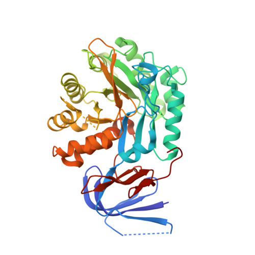
    The NA23_RS08100 gene of Fervidobacterium islandicum AW-1 encodes a keratin-degrading β-aspartyl peptidase ( Fi BAP) that is highly expressed under starvation conditions. Herein, we expressed the gene in Escherichia coli , purified the recombinant enzyme to homogeneity, and investigated its function. The 318 kDa recombinant Fi BAP enzyme exhibited maximal activity at 80°C and pH 7.0 in the presence of Zn 2+ . Size-exclusion chromatography revealed that the native enzyme is an octamer comprising a tetramer of dimers; this was further supported by determination of its crystal structure at 2.6 Å resolution. Consistently, the structure of Fi BAP revealed three additional salt bridges in each dimer, involving 12 ionic interactions that might contribute to its high thermostability. In addition, the co-crystal structure containing the substrate analog N -carbobenzoxy-β-Asp-Leu at 2.7 Å resolution revealed binuclear Zn 2+ -mediated substrate binding, suggesting that Fi BAP is a hyperthermophilic type-I IadA, in accordance with sequence-based phylogenetic analysis. Indeed, complementation of a Leu auxotrophic E. coli mutant strain (Δ iadA and Δ leuB ) with Fi BAP enabled the mutant strain to grow on isoAsp-Leu peptides. Remarkably, LC-MS/MS analysis of soluble keratin hydrolysates revealed that Fi BAP not only cleaves the C-terminus of isoAsp residues but also has a relatively broad substrate specificity toward α-peptide bonds. Moreover, heat shock-induced protein aggregates retarded bacterial growth, but expression of BAP alleviated the growth defect by degrading damaged proteins. Taken together, these results suggest that the viability of hyperthermophiles under stressful conditions may rely on the activity of BAP within cellular protein repair systems.


  • Organizational Affiliation
    • Department of Biotechnology, Yonsei University, Seoul, South Korea.

Macromolecule Content 

  • Total Structure Weight: 174.09 kDa 
  • Atom Count: 11,800 
  • Modeled Residue Count: 1,497 
  • Deposited Residue Count: 1,564 
  • Unique protein chains: 1

Macromolecules

Find similar proteins by:|  3D Structure
Entity ID: 1
MoleculeChains  Sequence LengthOrganismDetailsImage
Isoaspartyl dipeptidaseA [auth B],
B [auth A],
C,
D
391Fervidobacterium islandicumMutation(s): 0 
Gene Names: NA23_08080
EC: 3.4.19

Small Molecules

Ligands 4 Unique
IDChains Name / Formula / InChI Key2D Diagram3D Interactions
FWO
(Subject of Investigation/LOI)

Query on FWO


Download:Ideal Coordinates CCD File
AA [auth D],
I [auth B],
P [auth A],
W [auth C]
(2S)-2-[[(3S)-3-azanyl-4-oxidanyl-4-oxidanylidene-butanoyl]amino]-4-methyl-pentanoic acid
C10 H18 N2 O5
IYJILWQAFPUBHP-BQBZGAKWSA-N
PEG

Query on PEG


Download:Ideal Coordinates CCD File
M [auth A],
T [auth C]
DI(HYDROXYETHYL)ETHER
C4 H10 O3
MTHSVFCYNBDYFN-UHFFFAOYSA-N
GOL

Query on GOL


Download:Ideal Coordinates CCD File
E [auth B]
F [auth B]
G [auth B]
H [auth B]
L [auth A]
E [auth B],
F [auth B],
G [auth B],
H [auth B],
L [auth A],
N [auth A],
O [auth A],
S [auth C],
U [auth C],
V [auth C],
Z [auth D]
GLYCEROL
C3 H8 O3
PEDCQBHIVMGVHV-UHFFFAOYSA-N
ZN

Query on ZN


Download:Ideal Coordinates CCD File
BA [auth D]
CA [auth D]
J [auth B]
K [auth B]
Q [auth A]
BA [auth D],
CA [auth D],
J [auth B],
K [auth B],
Q [auth A],
R [auth A],
X [auth C],
Y [auth C]
ZINC ION
Zn
PTFCDOFLOPIGGS-UHFFFAOYSA-N

Experimental Data & Validation

Experimental Data

  • Method: X-RAY DIFFRACTION
  • Resolution: 2.75 Å
  • R-Value Free:  0.278 (Depositor), 0.275 (DCC) 
  • R-Value Work:  0.196 (Depositor), 0.197 (DCC) 
  • R-Value Observed: 0.200 (Depositor) 
Space Group: P 2 21 2
Unit Cell:
Length ( Å )Angle ( ˚ )
a = 78.609α = 90
b = 150.425β = 90
c = 151.996γ = 90
Software Package:
Software NamePurpose
HKL-2000data reduction
HKL-2000data scaling
REFMACrefinement
PDB_EXTRACTdata extraction
PHASERphasing

Structure Validation

View Full Validation Report


Ligand Structure Quality Assessment 


Entry History 

Deposition Data

Revision History  (Full details and data files)

  • Version 1.0: 2020-12-30
    Type: Initial release
  • Version 1.1: 2021-01-20
    Changes: Database references
  • Version 1.2: 2023-11-29
    Changes: Data collection, Database references, Refinement description