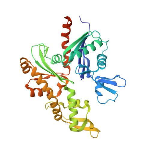
MreB5 Is a Determinant of Rod-to-Helical Transition in the Cell-Wall-less Bacterium Spiroplasma.
Harne, S., Duret, S., Pande, V., Bapat, M., Beven, L., Gayathri, P.(2020) Curr Biol 30: 4753
- PubMed: 32976813 Search on PubMed
- DOI: https://doi.org/10.1016/j.cub.2020.08.093
- Primary Citation Related Structures:
7BVY, 7BVZ - PubMed Abstract:
In most rod-shaped bacteria, the spatial coordination of cell wall synthesis machinery by MreBs is the main theme for shape determination and maintenance in cell-walled bacteria [1-9]. However, how rod or spiral shapes are achieved and maintained in cell-wall-less bacteria is currently unknown. Spiroplasma, a helical Mollicute that lacks cell wall synthesis genes, encodes five MreB paralogs and a unique cytoskeletal protein fibril [10, 11]. Here, we show that MreB5, one of the five MreB paralogs, contributes to cell elongation and is essential for the transition from rod-to-helical shape in Spiroplasma. Comparative genomic and proteomic characterization of a helical and motile wild-type Spiroplasma strain and a non-helical, non-motile natural variant helped delineate the specific roles of MreB5. Moreover, complementation of the non-helical strain with MreB5 restored its helical shape and motility by a kink-based mechanism described for Spiroplasma [12]. Earlier studies had proposed that length changes in fibril filaments are responsible for the change in handedness of the helical cell and kink propagation during motility [13]. Through structural and biochemical characterization, we identify that MreB5 exists as antiparallel double protofilaments that interact with fibril and the membrane, and thus potentially assists in kink propagation. In summary, our study provides direct experimental evidence for MreB in maintaining cell length, helical shape, and motility-revealing the role of MreB in sculpting the cell in the absence of a cell wall.
- Indian Institute of Science Education and Research, Dr. Homi Bhabha Road, Pashan, Pune 411008, India.
Organizational Affiliation: