7BP2 | pdb_00007bp2

Structural mechanism directing nucleosome reorganization by NAP1-RELATED PROTEIN 1 (NRP1)


Experimental Data Snapshot

  • Method: X-RAY DIFFRACTION
  • Resolution: 1.58 Å
  • R-Value Free: 
    0.186 (Depositor), 0.194 (DCC) 
  • R-Value Work: 
    0.155 (Depositor), 0.168 (DCC) 
  • R-Value Observed: 
    0.156 (Depositor) 

Starting Model: experimental
View more details

wwPDB Validation 3D Report Full Report

Validation slider image for 7BP2

Ligand Structure Quality Assessment 


This is version 1.4 of the entry. See complete history

Literature

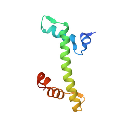
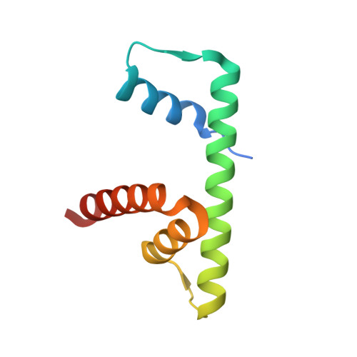
NAP1-Related Protein 1 (NRP1) has multiple interaction modes for chaperoning histones H2A-H2B.

Luo, Q.Wang, B.Wu, Z.Jiang, W.Wang, Y.Du, K.Zhou, N.Zheng, L.Gan, J.Shen, W.H.Ma, J.Dong, A.

(2020) Proc Natl Acad Sci U S A 117: 30391-30399

  • DOI: https://doi.org/10.1073/pnas.2011089117
  • Primary Citation Related Structures: 
    7BP2, 7BP4, 7BP5, 7BP6, 7C7X

  • PubMed Abstract: 

    Nucleosome Assembly Protein 1 (NAP1) family proteins are evolutionarily conserved histone chaperones that play important roles in diverse biological processes. In this study, we determined the crystal structure of Arabidopsis NAP1-Related Protein 1 (NRP1) complexed with H2A-H2B and uncovered a previously unknown interaction mechanism in histone chaperoning. Both in vitro binding and in vivo plant rescue assays proved that interaction mediated by the N-terminal α-helix (αN) domain is essential for NRP1 function. In addition, the C-terminal acidic domain (CTAD) of NRP1 binds to H2A-H2B through a conserved mode similar to other histone chaperones. We further extended previous knowledge of the NAP1-conserved earmuff domain by mapping the amino acids of NRP1 involved in association with H2A-H2B. Finally, we showed that H2A-H2B interactions mediated by αN, earmuff, and CTAD domains are all required for the effective chaperone activity of NRP1. Collectively, our results reveal multiple interaction modes of a NAP1 family histone chaperone and shed light on how histone chaperones shield H2A-H2B from nonspecific interaction with DNA.


  • Organizational Affiliation
    • State Key Laboratory of Genetic Engineering, Collaborative Innovation Center of Genetics and Development, International Associated Laboratory of CNRS-Fudan-HUNAU on Plant Epigenome Research, Department of Biochemistry, Institute of Plant Biology, School of Life Sciences, Fudan University, 200438 Shanghai, China.

Macromolecule Content 

  • Total Structure Weight: 43.46 kDa 
  • Atom Count: 3,188 
  • Modeled Residue Count: 352 
  • Deposited Residue Count: 382 
  • Unique protein chains: 2

Macromolecules

Find similar proteins by:|  3D Structure
Entity ID: 1
MoleculeChains  Sequence LengthOrganismDetailsImage
Histone H2A.6
A, C
93Arabidopsis thalianaMutation(s): 0 
Gene Names: RAT5H2A-1At5g54640MRB17.14
UniProt
Find proteins for Q9LD28 (Arabidopsis thaliana)
Explore Q9LD28 
Go to UniProtKB:  Q9LD28
Entity Groups
Sequence Clusters30% Identity50% Identity70% Identity90% Identity95% Identity100% Identity
UniProt GroupQ9LD28
Sequence Annotations
Expand
Reference Sequence
Find similar proteins by:|  3D Structure
Entity ID: 2
MoleculeChains  Sequence LengthOrganismDetailsImage
Histone H2B.1
B, D
98Arabidopsis thalianaMutation(s): 0 
Gene Names: At1g07790F24B9.10
UniProt
Find proteins for Q9LQQ4 (Arabidopsis thaliana)
Explore Q9LQQ4 
Go to UniProtKB:  Q9LQQ4
Entity Groups
Sequence Clusters30% Identity50% Identity70% Identity90% Identity95% Identity100% Identity
UniProt GroupQ9LQQ4
Sequence Annotations
Expand
Reference Sequence

Small Molecules

Ligands 2 Unique
IDChains Name / Formula / InChI Key2D Diagram3D Interactions
SO4
(Subject of Investigation/LOI)

Query on SO4


Download:Ideal Coordinates CCD File
E [auth A]
F [auth A]
H [auth B]
I [auth B]
J [auth B]
E [auth A],
F [auth A],
H [auth B],
I [auth B],
J [auth B],
L [auth C],
M [auth C],
N [auth D],
O [auth D],
P [auth D]
SULFATE ION
O4 S
QAOWNCQODCNURD-UHFFFAOYSA-L
GOL
(Subject of Investigation/LOI)

Query on GOL


Download:Ideal Coordinates CCD File
G [auth A],
K [auth B],
Q [auth D],
R [auth D]
GLYCEROL
C3 H8 O3
PEDCQBHIVMGVHV-UHFFFAOYSA-N

Experimental Data & Validation

Experimental Data

  • Method: X-RAY DIFFRACTION
  • Resolution: 1.58 Å
  • R-Value Free:  0.186 (Depositor), 0.194 (DCC) 
  • R-Value Work:  0.155 (Depositor), 0.168 (DCC) 
  • R-Value Observed: 0.156 (Depositor) 
Space Group: P 31
Unit Cell:
Length ( Å )Angle ( ˚ )
a = 65.712α = 90
b = 65.712β = 90
c = 96.687γ = 120
Software Package:
Software NamePurpose
HKL-2000data scaling
REFMACrefinement
PDB_EXTRACTdata extraction
HKL-2000data reduction
REFMACphasing

Structure Validation

View Full Validation Report


Ligand Structure Quality Assessment 


Entry History 

Deposition Data

Revision History  (Full details and data files)

  • Version 1.0: 2020-11-11
    Type: Initial release
  • Version 1.1: 2020-11-25
    Changes: Database references
  • Version 1.2: 2020-12-02
    Changes: Database references
  • Version 1.3: 2020-12-16
    Changes: Database references
  • Version 1.4: 2023-11-29
    Changes: Data collection, Database references, Refinement description