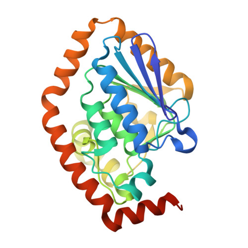
Structural and molecular dynamic studies of Pseudomonas aeruginosa OdaA reveal the regulation role of a C-terminal hinge element.
Zhao, N.L., Zhang, Q.Q., Zhao, C., Liu, L., Li, T., Li, C.C., He, L.H., Zhu, Y.B., Song, Y.J., Liu, H.X., Bao, R.(2020) Biochim Biophys Acta Gen Subj 1865: 129756-129756
- PubMed: 33010351 Search on PubMed
- DOI: https://doi.org/10.1016/j.bbagen.2020.129756
- Primary Citation Related Structures:
7BOR, 7CRD - PubMed Abstract:
Crotonase superfamily members exhibit great catalytic diversity towards various acyl-CoA substrates. A common CoA moiety binding pattern is usually observed in this family, understanding the substrate-binding mechanism would facilitate the rational engineering of crotonases for improved properties. We applied X-ray crystallography to investigate a putative enoyl-CoA hydratase/isomerase OdaA in Pseudomonas aeruginosa. Thermal shift assay (TSA) were performed to explore the binding of OdaA with CoA thioester substrates. Furthermore, we performed molecular dynamics (MD) simulations to elucidate the dynamics of its CoA-binding site. We solved the crystal structures of the apo and CoA-bound OdaA. Thermal shift assay (TSA) showed that CoA thioester substrates bind to OdaA with a different degree. MD simulations demonstrated that the C-terminal alpha helix underwent a structural transition and a hinge region would associate with this conformational change. TSA in combination with MD simulations elucidate that the dynamics of C-terminal alpha helix in CoA-binding, and a hinge region play an important role in conformational change. Those results help to extend our knowledge about the nature of crotonases and would be informative for future mechanistic studies and industry applications.
- Center of Infectious Diseases, State Key Laboratory of Biotherapy, West China Hospital, Sichuan University and Collaborative Innovation Center, Chengdu, China.
Organizational Affiliation: