7BNY | pdb_00007bny

Structure of 2A protein from encephalomyocarditis virus (EMCV)


Experimental Data Snapshot

  • Method: X-RAY DIFFRACTION
  • Resolution: 2.62 Å
  • R-Value Free: 
    0.251 (Depositor), 0.250 (DCC) 
  • R-Value Work: 
    0.225 (Depositor), 0.225 (DCC) 
  • R-Value Observed: 
    0.227 (Depositor) 

wwPDB Validation 3D Report Full Report

Validation slider image for 7BNY

This is version 1.2 of the entry. See complete history

Literature

Structural and molecular basis for Cardiovirus 2A protein as a viral gene expression switch.

Hill, C.H.Pekarek, L.Napthine, S.Kibe, A.Firth, A.E.Graham, S.C.Caliskan, N.Brierley, I.

(2021) Nat Commun 12: 7166-7166

  • DOI: https://doi.org/10.1038/s41467-021-27400-7
  • Primary Citation Related Structures: 
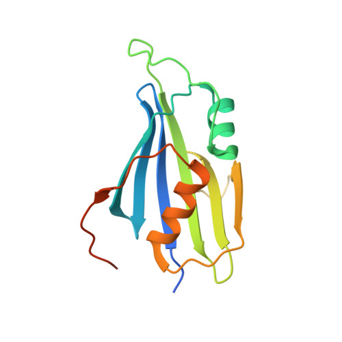
    7BNY

  • PubMed Abstract: 

    Programmed -1 ribosomal frameshifting (PRF) in cardioviruses is activated by the 2A protein, a multi-functional virulence factor that also inhibits cap-dependent translational initiation. Here we present the X-ray crystal structure of 2A and show that it selectively binds to a pseudoknot-like conformation of the PRF stimulatory RNA element in the viral genome. Using optical tweezers, we demonstrate that 2A stabilises this RNA element, likely explaining the increase in PRF efficiency in the presence of 2A. Next, we demonstrate a strong interaction between 2A and the small ribosomal subunit and present a cryo-EM structure of 2A bound to initiated 70S ribosomes. Multiple copies of 2A bind to the 16S rRNA where they may compete for binding with initiation and elongation factors. Together, these results define the structural basis for RNA recognition by 2A, show how 2A-mediated stabilisation of an RNA pseudoknot promotes PRF, and reveal how 2A accumulation may shut down translation during virus infection.


  • Organizational Affiliation
    • Division of Virology, Department of Pathology, University of Cambridge, Tennis Court Road, Cambridge, CB2 1QP, UK. chris.hill@york.ac.uk.

Macromolecule Content 

  • Total Structure Weight: 72.19 kDa 
  • Atom Count: 4,596 
  • Modeled Residue Count: 524 
  • Deposited Residue Count: 608 
  • Unique protein chains: 1

Macromolecules

Find similar proteins by:|  3D Structure
Entity ID: 1
MoleculeChains  Sequence LengthOrganismDetailsImage
Genome polyprotein
A, B, C, D
152Mengo virusMutation(s): 0 
EC: 3.6.4.13 (PDB Primary Data), 3.4.22.28 (PDB Primary Data), 2.7.7.48 (PDB Primary Data)
UniProt
Find proteins for P12296 (Mengo encephalomyocarditis virus)
Explore P12296 
Go to UniProtKB:  P12296
Entity Groups
Sequence Clusters30% Identity50% Identity70% Identity90% Identity95% Identity100% Identity
UniProt GroupP12296
Sequence Annotations
Expand
Reference Sequence

Small Molecules

Ligands 1 Unique
IDChains Name / Formula / InChI Key2D Diagram3D Interactions
SO4

Query on SO4


Download:Ideal Coordinates CCD File
E [auth A]
F [auth A]
G [auth A]
H [auth B]
I [auth B]
E [auth A],
F [auth A],
G [auth A],
H [auth B],
I [auth B],
J [auth B],
K [auth C],
L [auth D],
M [auth D]
SULFATE ION
O4 S
QAOWNCQODCNURD-UHFFFAOYSA-L

Experimental Data & Validation

Experimental Data

  • Method: X-RAY DIFFRACTION
  • Resolution: 2.62 Å
  • R-Value Free:  0.251 (Depositor), 0.250 (DCC) 
  • R-Value Work:  0.225 (Depositor), 0.225 (DCC) 
  • R-Value Observed: 0.227 (Depositor) 
Space Group: P 62 2 2
Unit Cell:
Length ( Å )Angle ( ˚ )
a = 91.56α = 90
b = 91.56β = 90
c = 316.39γ = 120
Software Package:
Software NamePurpose
GDAdata collection
xia2data reduction
XDSdata reduction
Aimlessdata scaling
SHARPphasing
SHELXDphasing
PHENIXrefinement
ARP/wARPmodel building
Cootmodel building

Structure Validation

View Full Validation Report


Entry History 

& Funding Information

Deposition Data


Funding OrganizationLocationGrant Number
Wellcome TrustUnited Kingdom202797/Z/16/Z
Wellcome TrustUnited Kingdom098406/Z/12/B
European Communitys Seventh Framework ProgrammeEuropean UnionBIOSTRUCT-X (Contract No. 283570)

Revision History  (Full details and data files)

  • Version 1.0: 2021-12-08
    Type: Initial release
  • Version 1.1: 2022-02-02
    Changes: Database references
  • Version 1.2: 2024-11-13
    Changes: Data collection, Structure summary