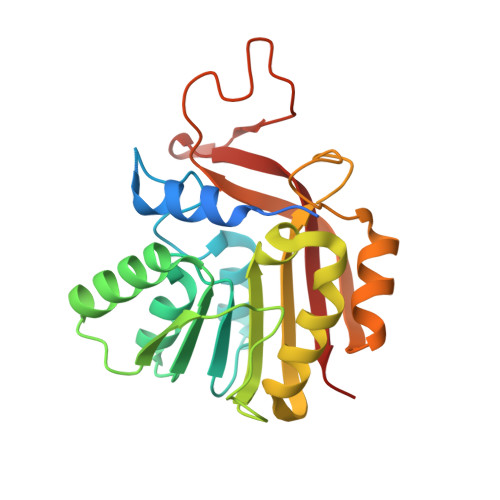
Structural basis of O-methylation of (2-heptyl-)1-hydroxyquinolin-4(1H)-one and related compounds by the heterocyclic toxin methyltransferase Rv0560c of Mycobacterium tuberculosis.
Sartor, P., Denkhaus, L., Gerhardt, S., Einsle, O., Fetzner, S.(2021) J Struct Biol 213: 107794-107794
- PubMed: 34506908
- DOI: https://doi.org/10.1016/j.jsb.2021.107794
- Primary Citation of Related Structures:
7BGG, 7NDM, 7NMK, 7NOY - PubMed Abstract:
The S-adenosyl-L-methionine-dependent methyltransferase Rv0560c of Mycobacterium tuberculosis belongs to an orthologous group of heterocyclic toxin methyltransferases (Htm) which likely contribute to resistance of mycobacteria towards antimicrobial natural compounds as well as drugs. Htm M.t. catalyzes the methylation of the Pseudomonas aeruginosa toxin 2-heptyl-1-hydroxyquinolin-4(1H)-one (also known as 2-heptyl-4-hydroxyquinoline N-oxide), a potent inhibitor of respiratory electron transfer, its 1-hydroxyquinolin-4(1H)-one core (QNO), structurally related (iso)quinolones, and some mycobactericidal compounds. In this study, crystal structures of Htm M.t. in complex with S-adenosyl-L-homocysteine (SAH) and the methyl-accepting substrates QNO or 4-hydroxyisoquinoline-1(2H)-one, or the methylated product 1-methoxyquinolin-4(1H)-one, were determined at < 1.9 Å resolution. The monomeric protein exhibits the typical Rossmann fold topology and conserved residues of class I methyltransferases. Its SAH binding pocket is connected via a short tunnel to a large solvent-accessible cavity, which accommodates the methyl-accepting substrate. Residues W44, F168, and F208 in connection with F212 form a hydrophobic clamp around the heteroaromatic ring of the methyl-accepting substrate and likely play a major role in substrate positioning. Structural and biochemical data suggest that H139 and T136 are key active site residues, with H139 acting as general base that activates the methyl-accepting hydroxy group. Our structural data may contribute to the design of Htm inhibitors or of antimycobacterial drugs unamenable for methylation.
- Institute of Molecular Microbiology and Biotechnology, University of Münster, Münster, Germany. Electronic address: sartor@uni-muenster.de.
Organizational Affiliation:


