7B43

Crystal structure of c-MET bound by compound 9


Experimental Data Snapshot

  • Method: X-RAY DIFFRACTION
  • Resolution: 1.87 Å
  • R-Value Free: 0.248 
  • R-Value Work: 0.200 
  • R-Value Observed: 0.203 

wwPDB Validation   3D Report Full Report


Ligand Structure Quality Assessment 


This is version 1.2 of the entry. See complete history


Literature

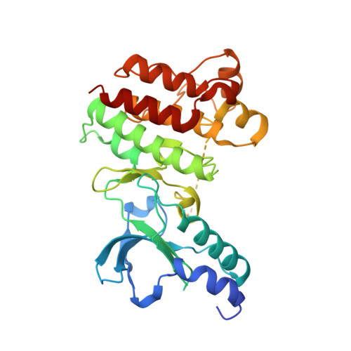
Structural Basis for Targeting the Folded P-Loop Conformation of c-MET.

Collie, G.W.Michaelides, I.N.Embrey, K.Stubbs, C.J.Borjesson, U.Dale, I.L.Snijder, A.Barlind, L.Song, K.Khurana, P.Phillips, C.Storer, R.I.

(2021) ACS Med Chem Lett 12: 162-167

  • DOI: https://doi.org/10.1021/acsmedchemlett.0c00392
  • Primary Citation of Related Structures:  
    7B3Q, 7B3T, 7B3V, 7B3W, 7B3Z, 7B40, 7B41, 7B42, 7B43, 7B44

  • PubMed Abstract: 

    We report here a fragment screen directed toward the c-MET kinase from which we discovered a series of inhibitors able to bind to a rare conformation of the protein in which the P-loop adopts a collapsed, or folded, arrangement. Preliminary SAR exploration led to an inhibitor ( 7 ) with nanomolar biochemical activity against c-MET and promising cell activity and kinase selectivity. These findings increase our structural understanding of the folded P-loop conformation of c-MET and provide a sound structural and chemical basis for further investigation of this underexplored yet potentially therapeutically exploitable conformational state.


  • Organizational Affiliation

    Discovery Sciences, R&D, AstraZeneca, Cambridge, United Kingdom.


Macromolecules
Find similar proteins by:  (by identity cutoff)  |  3D Structure
Entity ID: 1
MoleculeChains Sequence LengthOrganismDetailsImage
Hepatocyte growth factor receptor
A, B
299Homo sapiensMutation(s): 0 
Gene Names: MET
EC: 2.7.10.1
UniProt & NIH Common Fund Data Resources
Find proteins for P08581 (Homo sapiens)
Explore P08581 
Go to UniProtKB:  P08581
PHAROS:  P08581
GTEx:  ENSG00000105976 
Entity Groups  
Sequence Clusters30% Identity50% Identity70% Identity90% Identity95% Identity100% Identity
UniProt GroupP08581
Sequence Annotations
Expand
  • Reference Sequence
Small Molecules
Ligands 1 Unique
IDChains Name / Formula / InChI Key2D Diagram3D Interactions
SW5 (Subject of Investigation/LOI)
Query on SW5

Download Ideal Coordinates CCD File 
C [auth A],
D [auth B]
3-[(4-fluorophenyl)methyl]-5-(1-piperidin-4-ylpyrazol-4-yl)-1~{H}-pyrrolo[2,3-b]pyridine
C22 H22 F N5
YNHYKEIIJAKBHQ-UHFFFAOYSA-N
Experimental Data & Validation

Experimental Data

  • Method: X-RAY DIFFRACTION
  • Resolution: 1.87 Å
  • R-Value Free: 0.248 
  • R-Value Work: 0.200 
  • R-Value Observed: 0.203 
  • Space Group: P 1
Unit Cell:
Length ( Å )Angle ( ˚ )
a = 42.867α = 76.51
b = 59.738β = 73.88
c = 77.603γ = 69.25
Software Package:
Software NamePurpose
Aimlessdata scaling
BUSTERrefinement
PDB_EXTRACTdata extraction
XDSdata reduction
PHASERphasing

Structure Validation

View Full Validation Report


Ligand Structure Quality Assessment 


Entry History 

Deposition Data

  • Released Date: 2020-12-09 
  • Deposition Author(s): Collie, G.W.

Revision History  (Full details and data files)

  • Version 1.0: 2020-12-09
    Type: Initial release
  • Version 1.1: 2020-12-23
    Changes: Database references
  • Version 1.2: 2021-02-03
    Changes: Database references