7B2J | pdb_00007b2j

Crystal Structure of SARS-CoV-2 main protease (Nsp5) in complex with compound 5


Experimental Data Snapshot

  • Method: X-RAY DIFFRACTION
  • Resolution: 1.55 Å
  • R-Value Free: 
    0.203 (Depositor), 0.205 (DCC) 
  • R-Value Work: 
    0.162 (Depositor), 0.166 (DCC) 
  • R-Value Observed: 
    0.164 (Depositor) 

Starting Model: experimental
View more details

wwPDB Validation 3D Report Full Report

Validation slider image for 7B2J

Ligand Structure Quality Assessment 


This is version 1.2 of the entry. See complete history

Literature

Ultralarge Virtual Screening Identifies SARS-CoV-2 Main Protease Inhibitors with Broad-Spectrum Activity against Coronaviruses.

Luttens, A.Gullberg, H.Abdurakhmanov, E.Vo, D.D.Akaberi, D.Talibov, V.O.Nekhotiaeva, N.Vangeel, L.De Jonghe, S.Jochmans, D.Krambrich, J.Tas, A.Lundgren, B.Gravenfors, Y.Craig, A.J.Atilaw, Y.Sandstrom, A.Moodie, L.W.K.Lundkvist, A.van Hemert, M.J.Neyts, J.Lennerstrand, J.Kihlberg, J.Sandberg, K.Danielson, U.H.Carlsson, J.

(2022) J Am Chem Soc 144: 2905-2920

  • DOI: https://doi.org/10.1021/jacs.1c08402
  • Primary Citation Related Structures: 
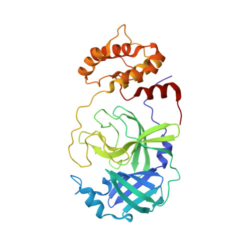
    7AU4, 7B2J, 7B2U, 7B5Z, 7B77, 7BIJ, 7NBT, 7NEO, 7O46, 7QBB

  • PubMed Abstract: 

    Drugs targeting SARS-CoV-2 could have saved millions of lives during the COVID-19 pandemic, and it is now crucial to develop inhibitors of coronavirus replication in preparation for future outbreaks. We explored two virtual screening strategies to find inhibitors of the SARS-CoV-2 main protease in ultralarge chemical libraries. First, structure-based docking was used to screen a diverse library of 235 million virtual compounds against the active site. One hundred top-ranked compounds were tested in binding and enzymatic assays. Second, a fragment discovered by crystallographic screening was optimized guided by docking of millions of elaborated molecules and experimental testing of 93 compounds. Three inhibitors were identified in the first library screen, and five of the selected fragment elaborations showed inhibitory effects. Crystal structures of target-inhibitor complexes confirmed docking predictions and guided hit-to-lead optimization, resulting in a noncovalent main protease inhibitor with nanomolar affinity, a promising in vitro pharmacokinetic profile, and broad-spectrum antiviral effect in infected cells.


  • Organizational Affiliation
    • Science for Life Laboratory, Department of Cell and Molecular Biology, Uppsala University, SE-75124 Uppsala, Sweden.

Macromolecule Content 

  • Total Structure Weight: 34.58 kDa 
  • Atom Count: 2,835 
  • Modeled Residue Count: 305 
  • Deposited Residue Count: 306 
  • Unique protein chains: 1

Macromolecules

Find similar proteins by:|  3D Structure
Entity ID: 1
MoleculeChains  Sequence LengthOrganismDetailsImage
3C-like proteinase nsp5306Severe acute respiratory syndrome coronavirus 2Mutation(s): 0 
Gene Names: rep1a-1b
EC: 3.4.22.69
UniProt
Find proteins for P0DTD1 (Severe acute respiratory syndrome coronavirus 2)
Explore P0DTD1 
Go to UniProtKB:  P0DTD1
Entity Groups
Sequence Clusters30% Identity50% Identity70% Identity90% Identity95% Identity100% Identity
UniProt GroupP0DTD1
Sequence Annotations
Expand
Reference Sequence

Small Molecules

Ligands 3 Unique
IDChains Name / Formula / InChI Key2D Diagram3D Interactions
SQ2
(Subject of Investigation/LOI)

Query on SQ2


Download:Ideal Coordinates CCD File
B [auth A]2-(1H-1,2,3-benzotriazol-1-yl)-1-(4-methylpiperidin-1-yl)ethan-1-one
C14 H18 N4 O
CTUYCKVPYLPOTM-UHFFFAOYSA-N
PEG

Query on PEG


Download:Ideal Coordinates CCD File
C [auth A]DI(HYDROXYETHYL)ETHER
C4 H10 O3
MTHSVFCYNBDYFN-UHFFFAOYSA-N
DMS

Query on DMS


Download:Ideal Coordinates CCD File
D [auth A],
E [auth A],
F [auth A],
G [auth A],
H [auth A]
DIMETHYL SULFOXIDE
C2 H6 O S
IAZDPXIOMUYVGZ-UHFFFAOYSA-N

Experimental Data & Validation

Experimental Data

  • Method: X-RAY DIFFRACTION
  • Resolution: 1.55 Å
  • R-Value Free:  0.203 (Depositor), 0.205 (DCC) 
  • R-Value Work:  0.162 (Depositor), 0.166 (DCC) 
  • R-Value Observed: 0.164 (Depositor) 
Space Group: C 1 2 1
Unit Cell:
Length ( Å )Angle ( ˚ )
a = 114.548α = 90
b = 53.652β = 100.559
c = 44.568γ = 90
Software Package:
Software NamePurpose
REFMACrefinement
PHENIXrefinement
PHASERphasing
Aimlessdata scaling
XDSdata reduction
Cootmodel building

Structure Validation

View Full Validation Report


Ligand Structure Quality Assessment 


Entry History 

Deposition Data

Revision History  (Full details and data files)

  • Version 1.0: 2022-02-23
    Type: Initial release
  • Version 1.1: 2022-03-02
    Changes: Database references
  • Version 1.2: 2024-01-31
    Changes: Data collection, Refinement description