7B2H

Crystal structure of the methyl-coenzyme M reductase from Methanothermobacter Marburgensis derivatized with xenon


Experimental Data Snapshot

  • Method: X-RAY DIFFRACTION
  • Resolution: 2.12 Å
  • R-Value Free: 0.204 
  • R-Value Work: 0.177 
  • R-Value Observed: 0.179 

wwPDB Validation   3D Report Full Report


This is version 1.1 of the entry. See complete history


Literature

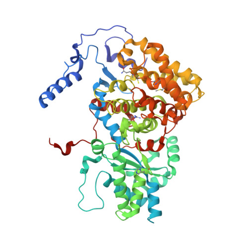
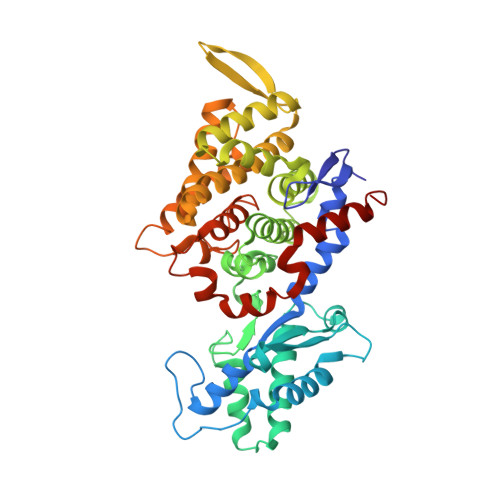
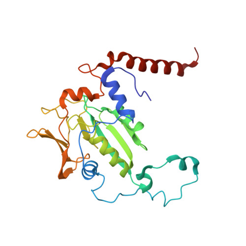
Crystal structure of a key enzyme for anaerobic ethane activation.

Hahn, C.J.Lemaire, O.N.Kahnt, J.Engilberge, S.Wegener, G.Wagner, T.

(2021) Science 373: 118-121

  • DOI: https://doi.org/10.1126/science.abg1765
  • Primary Citation of Related Structures:  
    7B1S, 7B2C, 7B2H

  • PubMed Abstract: 

    Ethane, the second most abundant hydrocarbon gas in the seafloor, is efficiently oxidized by anaerobic archaea in syntrophy with sulfate-reducing bacteria. Here, we report the 0.99-angstrom-resolution structure of the proposed ethane-activating enzyme and describe the specific traits that distinguish it from methane-generating and -consuming methyl-coenzyme M reductases. The widened catalytic chamber, harboring a dimethylated nickel-containing F 430 cofactor, would adapt the chemistry of methyl-coenzyme M reductases for a two-carbon substrate. A sulfur from methionine replaces the oxygen from a canonical glutamine as the nickel lower-axial ligand, a feature conserved in thermophilic ethanotrophs. Specific loop extensions, a four-helix bundle dilatation, and posttranslational methylations result in the formation of a 33-angstrom-long hydrophobic tunnel, which guides the ethane to the buried active site as confirmed with xenon pressurization experiments.


  • Organizational Affiliation

    Max Planck Institute for Marine Microbiology, Bremen 28359, Germany.


Macromolecules
Find similar proteins by:  (by identity cutoff)  |  3D Structure
Entity ID: 1
MoleculeChains Sequence LengthOrganismDetailsImage
Methyl-coenzyme M reductase I subunit alpha
A, D
550Methanothermobacter marburgensis str. MarburgMutation(s): 0 
EC: 2.8.4.1
UniProt
Find proteins for P11558 (Methanothermobacter marburgensis (strain ATCC BAA-927 / DSM 2133 / JCM 14651 / NBRC 100331 / OCM 82 / Marburg))
Explore P11558 
Go to UniProtKB:  P11558
Entity Groups  
Sequence Clusters30% Identity50% Identity70% Identity90% Identity95% Identity100% Identity
UniProt GroupP11558
Sequence Annotations
Expand
  • Reference Sequence
Find similar proteins by:  (by identity cutoff)  |  3D Structure
Entity ID: 2
MoleculeChains Sequence LengthOrganismDetailsImage
Methyl-coenzyme M reductase I subunit beta
B, E
443Methanothermobacter marburgensis str. MarburgMutation(s): 0 
EC: 2.8.4.1
UniProt
Find proteins for P11560 (Methanothermobacter marburgensis (strain ATCC BAA-927 / DSM 2133 / JCM 14651 / NBRC 100331 / OCM 82 / Marburg))
Explore P11560 
Go to UniProtKB:  P11560
Entity Groups  
Sequence Clusters30% Identity50% Identity70% Identity90% Identity95% Identity100% Identity
UniProt GroupP11560
Sequence Annotations
Expand
  • Reference Sequence
Find similar proteins by:  (by identity cutoff)  |  3D Structure
Entity ID: 3
MoleculeChains Sequence LengthOrganismDetailsImage
Methyl-coenzyme M reductase I subunit gamma
C, F
249Methanothermobacter marburgensis str. MarburgMutation(s): 0 
EC: 2.8.4.1
UniProt
Find proteins for P11562 (Methanothermobacter marburgensis (strain ATCC BAA-927 / DSM 2133 / JCM 14651 / NBRC 100331 / OCM 82 / Marburg))
Explore P11562 
Go to UniProtKB:  P11562
Entity Groups  
Sequence Clusters30% Identity50% Identity70% Identity90% Identity95% Identity100% Identity
UniProt GroupP11562
Sequence Annotations
Expand
  • Reference Sequence
Small Molecules
Ligands 11 Unique
IDChains Name / Formula / InChI Key2D Diagram3D Interactions
F43
Query on F43

Download Ideal Coordinates CCD File 
IA [auth D],
N [auth A]
FACTOR 430
C42 H51 N6 Ni O13
XLFIRMYGVLUNOY-SXMZNAGASA-M
TP7
Query on TP7

Download Ideal Coordinates CCD File 
H [auth A],
KA [auth D]
Coenzyme B
C11 H22 N O7 P S
JBJSVEVEEGOEBZ-SCZZXKLOSA-N
COM
Query on COM

Download Ideal Coordinates CCD File 
G [auth A],
JA [auth D]
1-THIOETHANESULFONIC ACID
C2 H6 O3 S2
ZNEWHQLOPFWXOF-UHFFFAOYSA-N
XE (Subject of Investigation/LOI)
Query on XE

Download Ideal Coordinates CCD File 
AB [auth E]
BA [auth B]
CA [auth B]
DA [auth B]
EA [auth B]
AB [auth E],
BA [auth B],
CA [auth B],
DA [auth B],
EA [auth B],
EB [auth F],
FA [auth B],
QA [auth D],
S [auth A],
XA [auth E],
YA [auth E],
ZA [auth E]
XENON
Xe
FHNFHKCVQCLJFQ-UHFFFAOYSA-N
PEG
Query on PEG

Download Ideal Coordinates CCD File 
DB [auth F]
J [auth A]
K [auth A]
MA [auth D]
NA [auth D]
DB [auth F],
J [auth A],
K [auth A],
MA [auth D],
NA [auth D],
OA [auth D],
TA [auth E],
W [auth B],
X [auth B]
DI(HYDROXYETHYL)ETHER
C4 H10 O3
MTHSVFCYNBDYFN-UHFFFAOYSA-N
SO4
Query on SO4

Download Ideal Coordinates CCD File 
T [auth A]SULFATE ION
O4 S
QAOWNCQODCNURD-UHFFFAOYSA-L
GOL
Query on GOL

Download Ideal Coordinates CCD File 
L [auth A]GLYCEROL
C3 H8 O3
PEDCQBHIVMGVHV-UHFFFAOYSA-N
EDO
Query on EDO

Download Ideal Coordinates CCD File 
M [auth A]1,2-ETHANEDIOL
C2 H6 O2
LYCAIKOWRPUZTN-UHFFFAOYSA-N
K
Query on K

Download Ideal Coordinates CCD File 
HA [auth B],
I [auth A],
RA [auth D],
U [auth A]
POTASSIUM ION
K
NPYPAHLBTDXSSS-UHFFFAOYSA-N
CL
Query on CL

Download Ideal Coordinates CCD File 
BB [auth E],
GA [auth B]
CHLORIDE ION
Cl
VEXZGXHMUGYJMC-UHFFFAOYSA-M
MG
Query on MG

Download Ideal Coordinates CCD File 
AA [auth B]
CB [auth F]
LA [auth D]
O [auth A]
P [auth A]
AA [auth B],
CB [auth F],
LA [auth D],
O [auth A],
P [auth A],
PA [auth D],
Q [auth A],
R [auth A],
SA [auth E],
UA [auth E],
V [auth B],
VA [auth E],
WA [auth E],
Y [auth B],
Z [auth B]
MAGNESIUM ION
Mg
JLVVSXFLKOJNIY-UHFFFAOYSA-N
Modified Residues  6 Unique
IDChains TypeFormula2D DiagramParent
AGM
Query on AGM
A, D
L-PEPTIDE LINKINGC7 H17 N4 O2ARG
DYA
Query on DYA
A, D
L-PEPTIDE LINKINGC4 H5 N O4ASP
GL3
Query on GL3
A, D
L-PEPTIDE LINKINGC2 H5 N O SGLY
MGN
Query on MGN
A, D
L-PEPTIDE LINKINGC6 H12 N2 O3GLN
MHS
Query on MHS
A, D
L-PEPTIDE LINKINGC7 H11 N3 O2HIS
SMC
Query on SMC
A, D
L-PEPTIDE LINKINGC4 H9 N O2 SCYS
Experimental Data & Validation

Experimental Data

  • Method: X-RAY DIFFRACTION
  • Resolution: 2.12 Å
  • R-Value Free: 0.204 
  • R-Value Work: 0.177 
  • R-Value Observed: 0.179 
  • Space Group: P 1 21 1
Unit Cell:
Length ( Å )Angle ( ˚ )
a = 81.719α = 90
b = 116.386β = 92.36
c = 123.07γ = 90
Software Package:
Software NamePurpose
PHENIXrefinement
XDSdata reduction
Aimlessdata scaling
PHASERphasing
PDB_EXTRACTdata extraction

Structure Validation

View Full Validation Report


Entry History & Funding Information

Deposition Data


Funding OrganizationLocationGrant Number
Max Planck SocietyGermany--
German Research Foundation (DFG)GermanyEXC-2077 390741603

Revision History  (Full details and data files)

  • Version 1.0: 2021-07-14
    Type: Initial release
  • Version 1.1: 2024-01-31
    Changes: Data collection, Database references, Derived calculations, Refinement description