


Crystal structures of phosphatidyl serine synthase PSS reveal the catalytic mechanism of CDP-DAG alcohol O-phosphatidyl transferases
Centola, M., Betz, H., Yildiz, O.(2021) Nat Commun 12: 6982
Experimental Data Snapshot
(2021) Nat Commun 12: 6982
Entity ID: 1 | |||||
|---|---|---|---|---|---|
| Molecule | Chains | Sequence Length | Organism | Details | Image |
| CDP-diacylglycerol--serine O-phosphatidyltransferase | 225 | Methanocaldococcus jannaschii DSM 2661 | Mutation(s): 0 Gene Names: pssA, MJ1212 EC: 2.7.8.8 Membrane Entity: Yes | 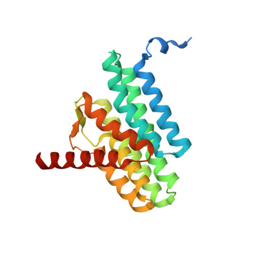 | |
UniProt | |||||
Entity Groups | |||||
| Sequence Clusters | 30% Identity50% Identity70% Identity90% Identity95% Identity100% Identity | ||||
| UniProt Group | Q58609 | ||||
Sequence AnnotationsExpand | |||||
Reference Sequence | |||||
| Ligands 3 Unique | |||||
|---|---|---|---|---|---|
| ID | Chains | Name / Formula / InChI Key | 2D Diagram | 3D Interactions | |
| 58A Download:Ideal Coordinates CCD File | C [auth A], G [auth B] | 5'-O-[(R)-{[(S)-{(2R)-2,3-bis[(9E)-octadec-9-enoyloxy]propoxy}(hydroxy)phosphoryl]oxy}(hydroxy)phosphoryl]cytidine C48 H85 N3 O15 P2 WVVFFOKRFKIBHD-ZIPNUMAKSA-N |  | ||
| CA Download:Ideal Coordinates CCD File | D [auth A], H [auth B] | CALCIUM ION Ca BHPQYMZQTOCNFJ-UHFFFAOYSA-N |  | ||
| CL Download:Ideal Coordinates CCD File | E [auth A], F [auth A], I [auth B], J [auth B] | CHLORIDE ION Cl VEXZGXHMUGYJMC-UHFFFAOYSA-M |  | ||
| Length ( Å ) | Angle ( ˚ ) |
|---|---|
| a = 62.13 | α = 90 |
| b = 70.81 | β = 90 |
| c = 94.09 | γ = 90 |
| Software Name | Purpose |
|---|---|
| PHENIX | refinement |
| XDS | data reduction |
| XSCALE | data scaling |
| PHASER | phasing |


