Unraveling a Force-Generating Allosteric Pathway of Actomyosin Communication Associated with ADP and P i Release.
Franz, P., Ewert, W., Preller, M., Tsiavaliaris, G.(2020) Int J Mol Sci 22
- PubMed: 33374308 Search on PubMedSearch on PubMed Central
- DOI: https://doi.org/10.3390/ijms22010104
- Primary Citation Related Structures:
7B19, 7B1A - PubMed Abstract:
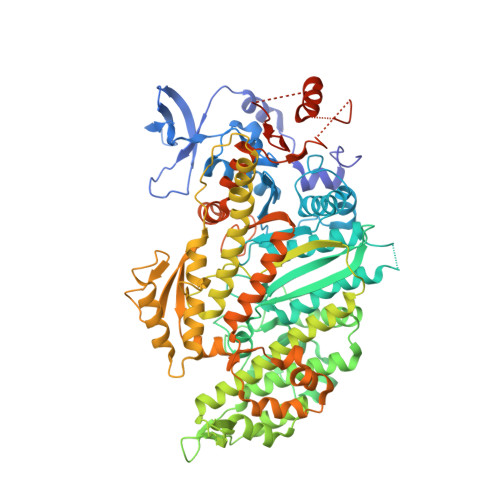
The actomyosin system generates mechanical work with the execution of the power stroke, an ATP-driven, two-step rotational swing of the myosin-neck that occurs post ATP hydrolysis during the transition from weakly to strongly actin-bound myosin states concomitant with P i release and prior to ADP dissociation. The activating role of actin on product release and force generation is well documented; however, the communication paths associated with weak-to-strong transitions are poorly characterized. With the aid of mutant analyses based on kinetic investigations and simulations, we identified the W-helix as an important hub coupling the structural changes of switch elements during ATP hydrolysis to temporally controlled interactions with actin that are passed to the central transducer and converter. Disturbing the W-helix/transducer pathway increased actin-activated ATP turnover and reduced motor performance as a consequence of prolonged duration of the strongly actin-attached states. Actin-triggered P i release was accelerated, while ADP release considerably decelerated, both limiting maximum ATPase, thus transforming myosin-2 into a high-duty-ratio motor. This kinetic signature of the mutant allowed us to define the fractional occupancies of intermediate states during the ATPase cycle providing evidence that myosin populates a cleft-closure state of strong actin interaction during the weak-to-strong transition with bound hydrolysis products before accomplishing the power stroke.
- Cellular Biophysics, Institute for Biophysical Chemistry, Hannover Medical School, 30625 Hannover, Germany.
Organizational Affiliation: