


Metadynamics simulations of CK2 compound unbinding to understand slow dissociation kinetics.
Date, M.To be published.
Experimental Data Snapshot
Entity ID: 1 | |||||
|---|---|---|---|---|---|
| Molecule | Chains | Sequence Length | Organism | Details | Image |
| Casein kinase II subunit alpha | 342 | Homo sapiens | Mutation(s): 0 Gene Names: CSNK2A1, CK2A1 EC: 2.7.11.1 | 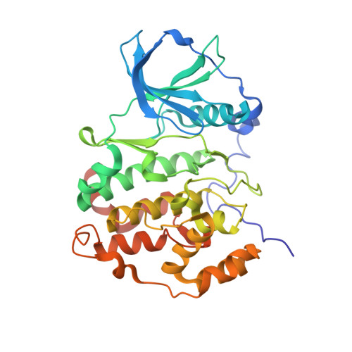 | |
UniProt & NIH Common Fund Data Resources | |||||
GTEx: ENSG00000101266 | |||||
Entity Groups | |||||
| Sequence Clusters | 30% Identity50% Identity70% Identity90% Identity95% Identity100% Identity | ||||
| UniProt Group | P68400 | ||||
Sequence AnnotationsExpand | |||||
Reference Sequence | |||||
| Ligands 3 Unique | |||||
|---|---|---|---|---|---|
| ID | Chains | Name / Formula / InChI Key | 2D Diagram | 3D Interactions | |
| S92 (Subject of Investigation/LOI) Download:Ideal Coordinates CCD File | I [auth A], Z [auth B] | 7-(cyclopropylamino)-5-(5-(6-oxo-1,6-dihydropyridin-3-yl)-1-(2-(piperidin-1-yl)ethyl)-1H-1,2,3-triazol-4-yl)pyrazolo[1,5-a]pyrimidine-3-carbonitrile C24 H26 N10 O DSBYVYTVSKXJPZ-UHFFFAOYSA-N |  | ||
| SO4 Download:Ideal Coordinates CCD File | C [auth A] D [auth A] E [auth A] F [auth A] G [auth A] | SULFATE ION O4 S QAOWNCQODCNURD-UHFFFAOYSA-L |  | ||
| EDO Download:Ideal Coordinates CCD File | AA [auth B] BA [auth B] CA [auth B] DA [auth B] EA [auth B] | 1,2-ETHANEDIOL C2 H6 O2 LYCAIKOWRPUZTN-UHFFFAOYSA-N |  | ||
| Length ( Å ) | Angle ( ˚ ) |
|---|---|
| a = 127.449 | α = 90 |
| b = 127.449 | β = 90 |
| c = 124.839 | γ = 90 |
| Software Name | Purpose |
|---|---|
| BUSTER | refinement |
| PDB_EXTRACT | data extraction |
| XDS | data reduction |
| SCALA | data scaling |
| AMoRE | phasing |


