7APU | pdb_00007apu

Structure of Adenylate kinase from Escherichia coli in complex with two ADP molecules refined at 1.36 A resolution.


Experimental Data Snapshot

  • Method: X-RAY DIFFRACTION
  • Resolution: 1.36 Å
  • R-Value Free: 
    0.206 (Depositor), 0.206 (DCC) 
  • R-Value Work: 
    0.184 (Depositor), 0.184 (DCC) 
  • R-Value Observed: 
    0.185 (Depositor) 

Starting Model: experimental
View more details

wwPDB Validation 3D Report Full Report

Validation slider image for 7APU

Ligand Structure Quality Assessment 


This is version 1.2 of the entry. See complete history

Literature

Dynamic Connection between Enzymatic Catalysis and Collective Protein Motions.

Ojeda-May, P.Mushtaq, A.U.Rogne, P.Verma, A.Ovchinnikov, V.Grundstrom, C.Dulko-Smith, B.Sauer, U.H.Wolf-Watz, M.Nam, K.

(2021) Biochemistry 60: 2246-2258

  • DOI: https://doi.org/10.1021/acs.biochem.1c00221
  • Primary Citation Related Structures: 
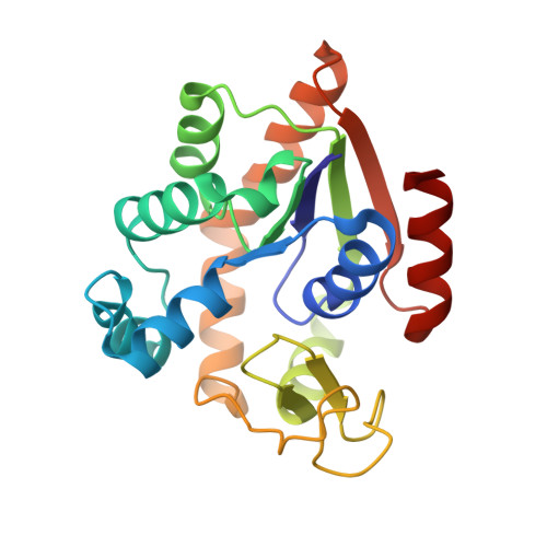
    7APU

  • PubMed Abstract: 

    Enzymes employ a wide range of protein motions to achieve efficient catalysis of chemical reactions. While the role of collective protein motions in substrate binding, product release, and regulation of enzymatic activity is generally understood, their roles in catalytic steps per se remain uncertain. Here, molecular dynamics simulations, enzyme kinetics, X-ray crystallography, and nuclear magnetic resonance spectroscopy are combined to elucidate the catalytic mechanism of adenylate kinase and to delineate the roles of catalytic residues in catalysis and the conformational change in the enzyme. This study reveals that the motions in the active site, which occur on a time scale of picoseconds to nanoseconds, link the catalytic reaction to the slow conformational dynamics of the enzyme by modulating the free energy landscapes of subdomain motions. In particular, substantial conformational rearrangement occurs in the active site following the catalytic reaction. This rearrangement not only affects the reaction barrier but also promotes a more open conformation of the enzyme after the reaction, which then results in an accelerated opening of the enzyme compared to that of the reactant state. The results illustrate a linkage between enzymatic catalysis and collective protein motions, whereby the disparate time scales between the two processes are bridged by a cascade of intermediate-scale motion of catalytic residues modulating the free energy landscapes of the catalytic and conformational change processes.


  • Organizational Affiliation
    • Department of Chemistry, Umeå University, Umeå SE-90187, Sweden.

Macromolecule Content 

  • Total Structure Weight: 48.99 kDa 
  • Atom Count: 4,090 
  • Modeled Residue Count: 428 
  • Deposited Residue Count: 428 
  • Unique protein chains: 1

Macromolecules

Find similar proteins by:|  3D Structure
Entity ID: 1
MoleculeChains  Sequence LengthOrganismDetailsImage
Adenylate kinase
A, B
214Escherichia coli K-12Mutation(s): 0 
Gene Names: adkdnaWplsAb0474JW0463
EC: 2.7.4.3
UniProt
Find proteins for P69441 (Escherichia coli (strain K12))
Explore P69441 
Go to UniProtKB:  P69441
Entity Groups
Sequence Clusters30% Identity50% Identity70% Identity90% Identity95% Identity100% Identity
UniProt GroupP69441
Sequence Annotations
Expand
Reference Sequence

Experimental Data & Validation

Experimental Data

  • Method: X-RAY DIFFRACTION
  • Resolution: 1.36 Å
  • R-Value Free:  0.206 (Depositor), 0.206 (DCC) 
  • R-Value Work:  0.184 (Depositor), 0.184 (DCC) 
  • R-Value Observed: 0.185 (Depositor) 
Space Group: P 21 21 2
Unit Cell:
Length ( Å )Angle ( ˚ )
a = 72.754α = 90
b = 82.226β = 90
c = 78.783γ = 90
Software Package:
Software NamePurpose
PHENIXrefinement
PHENIXrefinement
XDSdata reduction
Aimlessdata scaling
PHASERphasing
PDB_EXTRACTdata extraction

Structure Validation

View Full Validation Report


Ligand Structure Quality Assessment 


Entry History 

& Funding Information

Deposition Data


Funding OrganizationLocationGrant Number
Swedish Research CouncilSweden--

Revision History  (Full details and data files)

  • Version 1.0: 2022-03-02
    Type: Initial release
  • Version 1.1: 2022-12-21
    Changes: Database references
  • Version 1.2: 2024-01-31
    Changes: Data collection, Refinement description