7APL

tRNA-guanine transglycosylase G87C mutant spin-labeled with MTSL


Experimental Data Snapshot

  • Method: X-RAY DIFFRACTION
  • Resolution: 1.99 Å
  • R-Value Free: 0.209 
  • R-Value Work: 0.172 
  • R-Value Observed: 0.174 

wwPDB Validation   3D Report Full Report


This is version 1.3 of the entry. See complete history


Literature

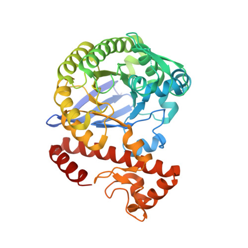
Unraveling a Ligand-Induced Twist of a Homodimeric Enzyme by Pulsed Electron-Electron Double Resonance.

Nguyen, D.Abdullin, D.Heubach, C.A.Pfaffeneder, T.Nguyen, A.Heine, A.Reuter, K.Diederich, F.Schiemann, O.Klebe, G.

(2021) Angew Chem Int Ed Engl 60: 23419-23426

  • DOI: https://doi.org/10.1002/anie.202108179
  • Primary Citation of Related Structures:  
    7APL, 7APM

  • PubMed Abstract: 

    Mechanistic insights into protein-ligand interactions can yield chemical tools for modulating protein function and enable their use for therapeutic purposes. For the homodimeric enzyme tRNA-guanine transglycosylase (TGT), a putative virulence target of shigellosis, ligand binding has been shown by crystallography to transform the functional dimer geometry into an incompetent twisted one. However, crystallographic observation of both end states does neither verify the ligand-induced transformation of one dimer into the other in solution nor does it shed light on the underlying transformation mechanism. We addressed these questions in an approach that combines site-directed spin labeling (SDSL) with distance measurements based on pulsed electron-electron double resonance (PELDOR or DEER) spectroscopy. We observed an equilibrium between the functional and twisted dimer that depends on the type of ligand, with a pyranose-substituted ligand being the most potent one in shifting the equilibrium toward the twisted dimer. Our experiments suggest a dissociation-association mechanism for the formation of the twisted dimer upon ligand binding.


  • Organizational Affiliation

    Institut für Pharmazeutische Chemie, Philipps-Universität Marburg, Marbacher Weg 8, 35032, Marburg, Germany.


Macromolecules
Find similar proteins by:  (by identity cutoff)  |  3D Structure
Entity ID: 1
MoleculeChains Sequence LengthOrganismDetailsImage
Queuine tRNA-ribosyltransferase388Zymomonas mobilis subsp. mobilis ZM4 = ATCC 31821Mutation(s): 4 
Gene Names: tgtZMO0363
EC: 2.4.2.29
UniProt
Find proteins for P28720 (Zymomonas mobilis subsp. mobilis (strain ATCC 31821 / ZM4 / CP4))
Explore P28720 
Go to UniProtKB:  P28720
Entity Groups  
Sequence Clusters30% Identity50% Identity70% Identity90% Identity95% Identity100% Identity
UniProt GroupP28720
Sequence Annotations
Expand
  • Reference Sequence
Small Molecules
Modified Residues  1 Unique
IDChains TypeFormula2D DiagramParent
R1A
Query on R1A
A
L-PEPTIDE LINKINGC12 H21 N2 O3 S2CYS
Experimental Data & Validation

Experimental Data

  • Method: X-RAY DIFFRACTION
  • Resolution: 1.99 Å
  • R-Value Free: 0.209 
  • R-Value Work: 0.172 
  • R-Value Observed: 0.174 
  • Space Group: C 2 2 21
Unit Cell:
Length ( Å )Angle ( ˚ )
a = 64.542α = 90
b = 90.947β = 90
c = 167.818γ = 90
Software Package:
Software NamePurpose
PHENIXrefinement
XDSdata reduction
XDSdata scaling
PHASERphasing
Cootmodel building

Structure Validation

View Full Validation Report


Entry History & Funding Information

Deposition Data


Funding OrganizationLocationGrant Number
German Research Foundation (DFG)Germany324043133

Revision History  (Full details and data files)

  • Version 1.0: 2020-10-28
    Type: Initial release
  • Version 1.1: 2021-10-06
    Changes: Data collection, Database references
  • Version 1.2: 2021-10-20
    Changes: Data collection, Database references
  • Version 1.3: 2024-01-31
    Changes: Data collection, Refinement description