7AOS

crystal structure of the RARalpha/RXRalpha ligand binding domain heterodimer in complex with a fragment of SRC1 coactivator


Experimental Data Snapshot

  • Method: X-RAY DIFFRACTION
  • Resolution: 2.55 Å
  • R-Value Free: 0.262 
  • R-Value Work: 0.205 
  • R-Value Observed: 0.211 

wwPDB Validation   3D Report Full Report


Ligand Structure Quality Assessment 


This is version 1.1 of the entry. See complete history


Literature

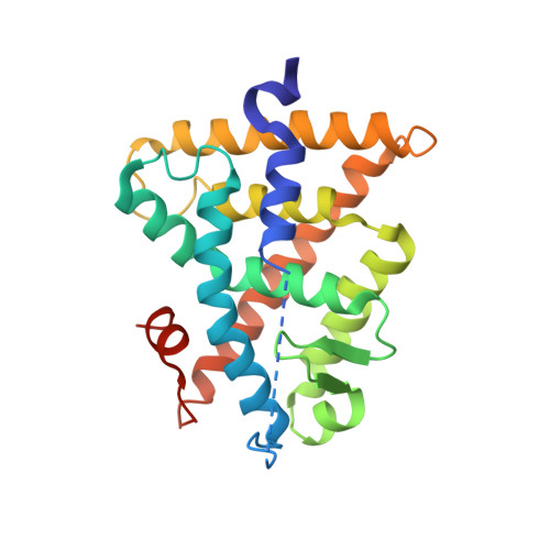
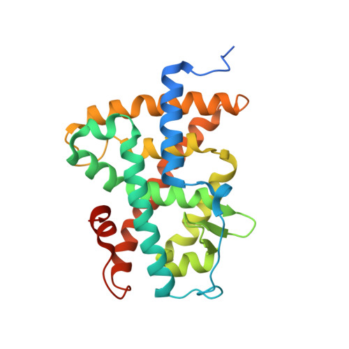
Structural Insights into the Interaction of the Intrinsically Disordered Co-activator TIF2 with Retinoic Acid Receptor Heterodimer (RXR/RAR).

Senicourt, L.le Maire, A.Allemand, F.Carvalho, J.E.Guee, L.Germain, P.Schubert, M.Bernado, P.Bourguet, W.Sibille, N.

(2021) J Mol Biol 433: 166899-166899

  • DOI: https://doi.org/10.1016/j.jmb.2021.166899
  • Primary Citation of Related Structures:  
    7AOS, 7APO, 7BK4

  • PubMed Abstract: 

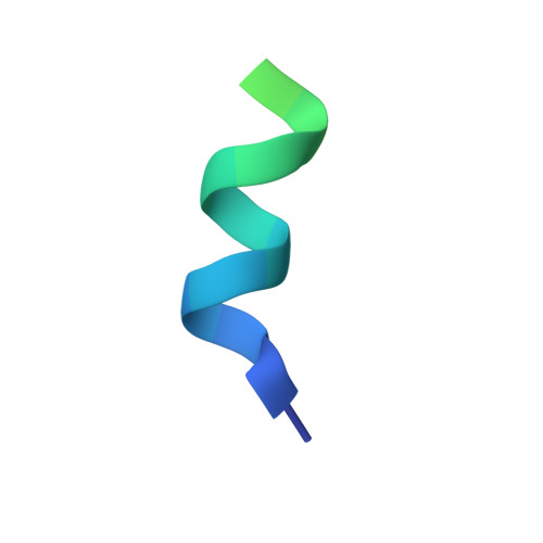
    Retinoic acid receptors (RARs) and retinoid X receptors (RXRs) form heterodimers that activate target gene transcription by recruiting co-activator complexes in response to ligand binding. The nuclear receptor (NR) co-activator TIF2 mediates this recruitment by interacting with the ligand-binding domain (LBD) of NRs trough the nuclear receptor interaction domain (TIF2 NRID ) containing three highly conserved α-helical LxxLL motifs (NR-boxes). The precise binding mode of this domain to RXR/RAR is not clear due to the disordered nature of TIF2. Here we present the structural characterization of TIF2 NRID by integrating several experimental (NMR, SAXS, Far-UV CD, SEC-MALS) and computational data. Collectively, the data are in agreement with a largely disordered protein with partially structured regions, including the NR-boxes and their flanking regions, which are evolutionary conserved. NMR and X-ray crystallographic data on TIF2 NRID in complex with RXR/RAR reveal a multisite binding of the three NR-boxes as well as an active role of their flanking regions in the interaction.


  • Organizational Affiliation

    Centre de Biochimie Structurale (CBS), CNRS, INSERM, Univ Montpellier, Montpellier, France.


Macromolecules
Find similar proteins by:  (by identity cutoff)  |  3D Structure
Entity ID: 1
MoleculeChains Sequence LengthOrganismDetailsImage
Retinoic acid receptor RXR-alpha238Mus musculusMutation(s): 0 
Gene Names: RxraNr2b1
UniProt
Find proteins for P28700 (Mus musculus)
Explore P28700 
Go to UniProtKB:  P28700
Entity Groups  
Sequence Clusters30% Identity50% Identity70% Identity90% Identity95% Identity100% Identity
UniProt GroupP28700
Sequence Annotations
Expand
  • Reference Sequence
Find similar proteins by:  (by identity cutoff)  |  3D Structure
Entity ID: 2
MoleculeChains Sequence LengthOrganismDetailsImage
Retinoic acid receptor alpha266Homo sapiensMutation(s): 0 
Gene Names: RARANR1B1
UniProt & NIH Common Fund Data Resources
Find proteins for P10276 (Homo sapiens)
Explore P10276 
Go to UniProtKB:  P10276
PHAROS:  P10276
GTEx:  ENSG00000131759 
Entity Groups  
Sequence Clusters30% Identity50% Identity70% Identity90% Identity95% Identity100% Identity
UniProt GroupP10276
Sequence Annotations
Expand
  • Reference Sequence
Find similar proteins by:  (by identity cutoff)  |  3D Structure
Entity ID: 3
MoleculeChains Sequence LengthOrganismDetailsImage
Nuclear receptor coactivator 1
C, D
27Homo sapiensMutation(s): 0 
EC: 2.3.1.48
UniProt & NIH Common Fund Data Resources
Find proteins for Q15788 (Homo sapiens)
Explore Q15788 
Go to UniProtKB:  Q15788
PHAROS:  Q15788
GTEx:  ENSG00000084676 
Entity Groups  
Sequence Clusters30% Identity50% Identity70% Identity90% Identity95% Identity100% Identity
UniProt GroupQ15788
Sequence Annotations
Expand
  • Reference Sequence
Small Molecules
Ligands 3 Unique
IDChains Name / Formula / InChI Key2D Diagram3D Interactions
LG2
Query on LG2

Download Ideal Coordinates CCD File 
E [auth A]6-[1-(3,5,5,8,8-PENTAMETHYL-5,6,7,8-TETRAHYDRONAPHTHALEN-2-YL)CYCLOPROPYL]PYRIDINE-3-CARBOXYLIC ACID
C24 H29 N O2
SLXTWXQUEZSSTJ-UHFFFAOYSA-N
EQN
Query on EQN

Download Ideal Coordinates CCD File 
G [auth B]4-{[(5,5,8,8-tetramethyl-5,6,7,8-tetrahydronaphthalen-2-yl)carbonyl]amino}benzoic acid
C22 H25 N O3
SZWKGOZKRMMLAJ-UHFFFAOYSA-N
GOL
Query on GOL

Download Ideal Coordinates CCD File 
F [auth A]GLYCEROL
C3 H8 O3
PEDCQBHIVMGVHV-UHFFFAOYSA-N
Experimental Data & Validation

Experimental Data

  • Method: X-RAY DIFFRACTION
  • Resolution: 2.55 Å
  • R-Value Free: 0.262 
  • R-Value Work: 0.205 
  • R-Value Observed: 0.211 
  • Space Group: P 43 21 2
Unit Cell:
Length ( Å )Angle ( ˚ )
a = 108.283α = 90
b = 108.283β = 90
c = 99.786γ = 90
Software Package:
Software NamePurpose
PHENIXrefinement
XDSdata reduction
PHASERphasing

Structure Validation

View Full Validation Report


Ligand Structure Quality Assessment 


Entry History & Funding Information

Deposition Data


Funding OrganizationLocationGrant Number
Brazilian National Council for Scientific and Technological Development (CNPq)Brazil--

Revision History  (Full details and data files)

  • Version 1.0: 2021-08-04
    Type: Initial release
  • Version 1.1: 2024-01-31
    Changes: Data collection, Database references, Refinement description