7AI7

MutS in Intermediate state


Experimental Data Snapshot

  • Method: ELECTRON MICROSCOPY
  • Resolution: 3.90 Å
  • Aggregation State: PARTICLE 
  • Reconstruction Method: SINGLE PARTICLE 

wwPDB Validation   3D Report Full Report


This is version 1.2 of the entry. See complete history


Literature

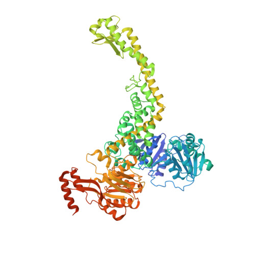
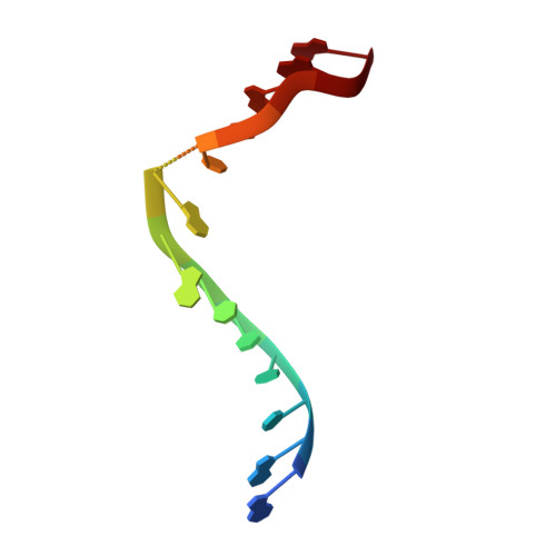
The selection process of licensing a DNA mismatch for repair.

Fernandez-Leiro, R.Bhairosing-Kok, D.Kunetsky, V.Laffeber, C.Winterwerp, H.H.Groothuizen, F.Fish, A.Lebbink, J.H.G.Friedhoff, P.Sixma, T.K.Lamers, M.H.

(2021) Nat Struct Mol Biol 28: 373-381

  • DOI: 10.1038/s41594-021-00577-7
  • Primary Citation of Related Structures:  
    7AI5, 7AI6, 7AI7, 7AIB, 7AIC

  • PubMed Abstract: 
  • DNA mismatch repair detects and removes mismatches from DNA by a conserved mechanism, reducing the error rate of DNA replication by 100- to 1,000-fold. In this process, MutS homologs scan DNA, recognize mismatches and initiate repair. How the MutS homologs selectively license repair of a mismatch among millions of matched base pairs is not understood ...

    DNA mismatch repair detects and removes mismatches from DNA by a conserved mechanism, reducing the error rate of DNA replication by 100- to 1,000-fold. In this process, MutS homologs scan DNA, recognize mismatches and initiate repair. How the MutS homologs selectively license repair of a mismatch among millions of matched base pairs is not understood. Here we present four cryo-EM structures of Escherichia coli MutS that provide snapshots, from scanning homoduplex DNA to mismatch binding and MutL activation via an intermediate state. During scanning, the homoduplex DNA forms a steric block that prevents MutS from transitioning into the MutL-bound clamp state, which can only be overcome through kinking of the DNA at a mismatch. Structural asymmetry in all four structures indicates a division of labor between the two MutS monomers. Together, these structures reveal how a small conformational change from the homoduplex- to heteroduplex-bound MutS acts as a licensing step that triggers a dramatic conformational change that enables MutL binding and initiation of the repair cascade.


    Organizational Affiliation

    Department of Cell and Chemical Biology, Leiden University Medical Center, Leiden, the Netherlands. m.h.lamers@lumc.nl.


Macromolecules

Find similar proteins by:  (by identity cutoff)  |  3D Structure
Entity ID: 1
MoleculeChainsSequence LengthOrganismDetailsImage
DNA mismatch repair protein MutSA, B853Escherichia coli K-12Mutation(s): 0 
Gene Names: mutSfdvb2733JW2703
UniProt
Find proteins for P23909 (Escherichia coli (strain K12))
Explore P23909 
Go to UniProtKB:  P23909
Entity Groups  
Sequence Clusters30% Identity50% Identity70% Identity90% Identity95% Identity100% Identity
UniProt GroupP23909
Protein Feature View
Expand
  • Reference Sequence

Find similar nucleic acids by:  Sequence   |   3D Structure  

Entity ID: 2
MoleculeChainsLengthOrganismImage
DNA (5'-D(P*GP*AP*TP*CP*CP*TP*AP*AP*CP*TP*AP*AP*G)-3')C14synthetic construct
Protein Feature View
Expand
  • Reference Sequence

Find similar nucleic acids by:  Sequence   |   3D Structure  

Entity ID: 3
MoleculeChainsLengthOrganismImage
DNA (5'-D(P*CP*TP*TP*AP*GP*CP*TP*TP*AP*GP*GP*AP*TP*C)-3')D14synthetic construct
Protein Feature View
Expand
  • Reference Sequence
Small Molecules
Ligands 1 Unique
IDChainsName / Formula / InChI Key2D Diagram3D Interactions
ADP
Query on ADP

Download Ideal Coordinates CCD File 
E [auth A],
F [auth B]
ADENOSINE-5'-DIPHOSPHATE
C10 H15 N5 O10 P2
XTWYTFMLZFPYCI-KQYNXXCUSA-N
 Ligand Interaction
Experimental Data & Validation

Experimental Data

  • Method: ELECTRON MICROSCOPY
  • Resolution: 3.90 Å
  • Aggregation State: PARTICLE 
  • Reconstruction Method: SINGLE PARTICLE 
EM Software:
TaskSoftware PackageVersion
MODEL REFINEMENTREFMAC5
RECONSTRUCTIONRELION3.1

Structure Validation

View Full Validation Report


Entry History & Funding Information

Deposition Data


Funding OrganizationLocationGrant Number
Medical Research Council (MRC, United Kingdom)United KingdomU105197143

Revision History  (Full details and data files)

  • Version 1.0: 2021-03-31
    Type: Initial release
  • Version 1.1: 2021-04-21
    Changes: Database references
  • Version 1.2: 2021-04-28
    Changes: Database references