7AC8

Molecular basis for the unique allosteric activation mechanism of the heterodimeric imidazole glycerol phosphate synthase complex.


Experimental Data Snapshot

  • Method: X-RAY DIFFRACTION
  • Resolution: 2.06 Å
  • R-Value Free: 0.186 
  • R-Value Work: 0.156 
  • R-Value Observed: 0.158 

wwPDB Validation   3D Report Full Report


Ligand Structure Quality Assessment 


This is version 1.2 of the entry. See complete history


Literature

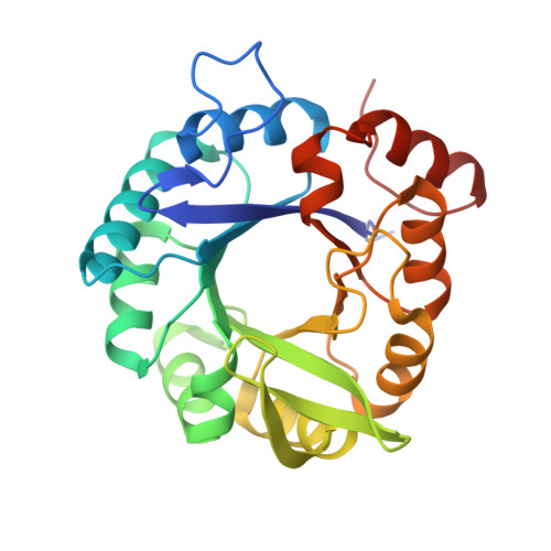
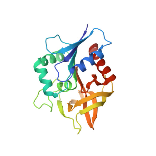
Molecular basis for the allosteric activation mechanism of the heterodimeric imidazole glycerol phosphate synthase complex.

Wurm, J.P.Sung, S.Kneuttinger, A.C.Hupfeld, E.Sterner, R.Wilmanns, M.Sprangers, R.

(2021) Nat Commun 12: 2748-2748

  • DOI: https://doi.org/10.1038/s41467-021-22968-6
  • Primary Citation of Related Structures:  
    7AC8

  • PubMed Abstract: 

    Imidazole glycerol phosphate synthase (HisFH) is a heterodimeric bienzyme complex operating at a central branch point of metabolism. HisFH is responsible for the HisH-catalyzed hydrolysis of glutamine to glutamate and ammonia, which is then used for a cyclase reaction by HisF. The HisFH complex is allosterically regulated but the underlying mechanism is not well understood. Here, we elucidate the molecular basis of the long range, allosteric activation of HisFH. We establish that the catalytically active HisFH conformation is only formed when the substrates of both HisH and HisF are bound. We show that in this conformation an oxyanion hole in the HisH active site is established, which rationalizes the observed 4500-fold allosteric activation compared to the inactive conformation. In solution, the inactive and active conformations are in a dynamic equilibrium and the HisFH turnover rates correlate with the population of the active conformation, which is in accordance with the ensemble model of allostery.


  • Organizational Affiliation

    Institute of Biophysics and Physical Biochemistry, Regensburg Center for Biochemistry, University of Regensburg, Regensburg, Germany.


Macromolecules
Find similar proteins by:  (by identity cutoff)  |  3D Structure
Entity ID: 1
MoleculeChains Sequence LengthOrganismDetailsImage
Imidazole glycerol phosphate synthase subunit HisF
A, C, E
253Thermotoga maritimaMutation(s): 0 
Gene Names: hisFTM_1036
EC: 4.3.2.10
UniProt
Find proteins for Q9X0C6 (Thermotoga maritima (strain ATCC 43589 / DSM 3109 / JCM 10099 / NBRC 100826 / MSB8))
Explore Q9X0C6 
Go to UniProtKB:  Q9X0C6
Entity Groups  
Sequence Clusters30% Identity50% Identity70% Identity90% Identity95% Identity100% Identity
UniProt GroupQ9X0C6
Sequence Annotations
Expand
  • Reference Sequence
Find similar proteins by:  (by identity cutoff)  |  3D Structure
Entity ID: 2
MoleculeChains Sequence LengthOrganismDetailsImage
Imidazole glycerol phosphate synthase subunit HisH
B, D, F
203Thermotoga maritima MSB8Mutation(s): 1 
Gene Names: hisHTM_1038
EC: 4.3.2.10 (PDB Primary Data), 3.5.1.2 (PDB Primary Data)
UniProt
Find proteins for Q9X0C8 (Thermotoga maritima (strain ATCC 43589 / DSM 3109 / JCM 10099 / NBRC 100826 / MSB8))
Explore Q9X0C8 
Go to UniProtKB:  Q9X0C8
Entity Groups  
Sequence Clusters30% Identity50% Identity70% Identity90% Identity95% Identity100% Identity
UniProt GroupQ9X0C8
Sequence Annotations
Expand
  • Reference Sequence
Small Molecules
Ligands 3 Unique
IDChains Name / Formula / InChI Key2D Diagram3D Interactions
GUO (Subject of Investigation/LOI)
Query on GUO

Download Ideal Coordinates CCD File 
G [auth A],
K [auth E]
[(2R,3S,4R,5R)-5-[4-aminocarbonyl-5-[(E)-[[(2R,3R,4S,5R)-3,4-bis(oxidanyl)-5-(phosphonooxymethyl)oxolan-2-yl]amino]methylideneamino]imidazol-1-yl]-3,4-bis(oxidanyl)oxolan-2-yl]methyl dihydrogen phosphate
C15 H25 N5 O15 P2
QOUSHGMTBIIAHR-KEOHHSTQSA-N
GLN
Query on GLN

Download Ideal Coordinates CCD File 
H [auth B],
J [auth D],
L [auth F]
GLUTAMINE
C5 H10 N2 O3
ZDXPYRJPNDTMRX-VKHMYHEASA-N
PO4 (Subject of Investigation/LOI)
Query on PO4

Download Ideal Coordinates CCD File 
I [auth C]PHOSPHATE ION
O4 P
NBIIXXVUZAFLBC-UHFFFAOYSA-K
Experimental Data & Validation

Experimental Data

  • Method: X-RAY DIFFRACTION
  • Resolution: 2.06 Å
  • R-Value Free: 0.186 
  • R-Value Work: 0.156 
  • R-Value Observed: 0.158 
  • Space Group: P 32
Unit Cell:
Length ( Å )Angle ( ˚ )
a = 92.835α = 90
b = 92.835β = 90
c = 168.625γ = 120
Software Package:
Software NamePurpose
PHENIXrefinement
XDSdata reduction
Aimlessdata scaling
PHASERphasing

Structure Validation

View Full Validation Report


Ligand Structure Quality Assessment 


Entry History & Funding Information

Deposition Data


Funding OrganizationLocationGrant Number
H2020 Marie Curie Actions of the European CommissionEuropean Union664726

Revision History  (Full details and data files)

  • Version 1.0: 2021-05-19
    Type: Initial release
  • Version 1.1: 2021-05-26
    Changes: Database references
  • Version 1.2: 2024-01-31
    Changes: Data collection, Database references, Refinement description