The molecular basis of spectral tuning in blue- and red-shifted flavin-binding fluorescent proteins.
Rollen, K., Granzin, J., Remeeva, A., Davari, M.D., Gensch, T., Nazarenko, V.V., Kovalev, K., Bogorodskiy, A., Borshchevskiy, V., Hemmer, S., Schwaneberg, U., Gordeliy, V., Jaeger, K.E., Batra-Safferling, R., Gushchin, I., Krauss, U.(2021) J Biol Chem 296: 100662-100662
- PubMed: 33862085 Search on PubMedSearch on PubMed Central
- DOI: https://doi.org/10.1016/j.jbc.2021.100662
- Primary Citation Related Structures:
6YX4, 6YX6, 6YXB, 7AB6, 7AB7, 7ABY - PubMed Abstract:
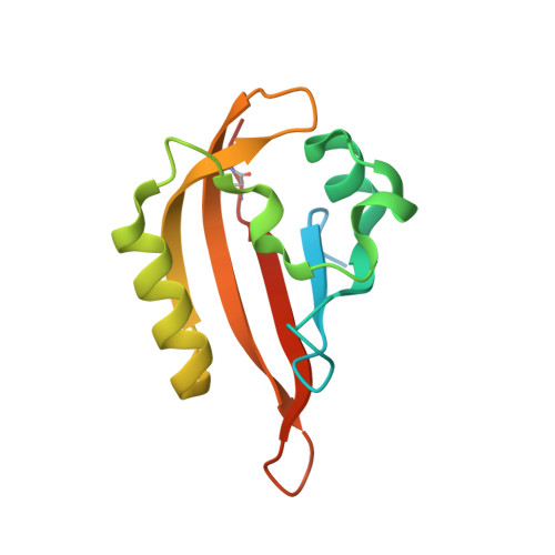
Photoactive biological systems modify the optical properties of their chromophores, known as spectral tuning. Determining the molecular origin of spectral tuning is instrumental for understanding the function and developing applications of these biomolecules. Spectral tuning in flavin-binding fluorescent proteins (FbFPs), an emerging class of fluorescent reporters, is limited by their dependency on protein-bound flavins, whose structure and hence electronic properties cannot be altered by mutation. A blue-shifted variant of the plant-derived improved light, oxygen, voltage FbFP has been created by introducing a lysine within the flavin-binding pocket, but the molecular basis of this shift remains unconfirmed. We here structurally characterize the blue-shifted improved light, oxygen, voltage variant and construct a new blue-shifted CagFbFP protein by introducing an analogous mutation. X-ray structures of both proteins reveal displacement of the lysine away from the chromophore and opening up of the structure as instrumental for the blue shift. Site saturation mutagenesis and high-throughput screening yielded a red-shifted variant, and structural analysis revealed that the lysine side chain of the blue-shifted variant is stabilized close to the flavin by a secondary mutation, accounting for the red shift. Thus, a single additional mutation in a blue-shifted variant is sufficient to generate a red-shifted FbFP. Using spectroscopy, X-ray crystallography, and quantum mechanics molecular mechanics calculations, we provide a firm structural and functional understanding of spectral tuning in FbFPs. We also show that the identified blue- and red-shifted variants allow for two-color microscopy based on spectral separation. In summary, the generated blue- and red-shifted variants represent promising new tools for application in life sciences.
- Institut für Molekulare Enzymtechnologie, Heinrich-Heine-Universität Düsseldorf, Forschungszentrum Jülich GmbH, Jülich, Germany.
Organizational Affiliation: