Development of a Protein Scaffold for Arginine Sensing Generated through the Dissection of the Arginine-Binding Protein from Thermotoga maritima .
Smaldone, G., Ruggiero, A., Balasco, N., Vitagliano, L.(2020) Int J Mol Sci 21
- PubMed: 33053818 Search on PubMedSearch on PubMed Central
- DOI: https://doi.org/10.3390/ijms21207503
- Primary Citation Related Structures:
7A99 - PubMed Abstract:
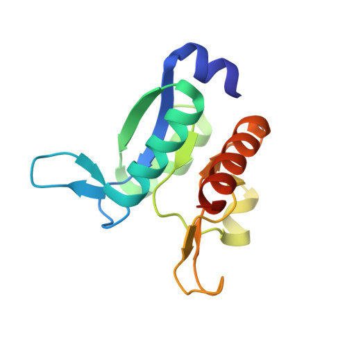
Arginine is one of the most important nutrients of living organisms as it plays a major role in important biological pathways. However, the accumulation of arginine as consequence of metabolic defects causes hyperargininemia, an autosomal recessive disorder. Therefore, the efficient detection of the arginine is a field of relevant biomedical/biotechnological interest. Here, we developed protein variants suitable for arginine sensing by mutating and dissecting the multimeric and multidomain structure of Thermotoga maritima arginine-binding protein (TmArgBP). Indeed, previous studies have shown that TmArgBP domain-swapped structure can be manipulated to generate simplified monomeric and single domain scaffolds. On both these stable scaffolds, to measure tryptophan fluorescence variations associated with the arginine binding, a Phe residue of the ligand binding pocket was mutated to Trp. Upon arginine binding, both mutants displayed a clear variation of the Trp fluorescence. Notably, the single domain scaffold variant exhibited a good affinity (~3 µM) for the ligand. Moreover, the arginine binding to this variant could be easily reverted under very mild conditions. Atomic-level data on the recognition process between the scaffold and the arginine were obtained through the determination of the crystal structure of the adduct. Collectively, present data indicate that TmArgBP scaffolds represent promising candidates for developing arginine biosensors.
- IRCCS SDN, Via Emanuele Gianturco, 113 80143 Naples, Italy.
Organizational Affiliation: