7A6Z

Structure of P226G BlaC from Mycobacterium tuberculosis


Experimental Data Snapshot

  • Method: X-RAY DIFFRACTION
  • Resolution: 1.30 Å
  • R-Value Free: 0.137 
  • R-Value Work: 0.116 
  • R-Value Observed: 0.117 

wwPDB Validation   3D Report Full Report


This is version 1.3 of the entry. See complete history


Literature

Conserved proline residues prevent dimerization and aggregation in the beta-lactamase BlaC.

Chikunova, A.Manley, M.P.Heijjer, C.N.Drenth, C.S.Cramer-Blok, A.J.Ahmad, M.U.D.Perrakis, A.Ubbink, M.

(2024) Protein Sci 33: e4972-e4972

  • DOI: https://doi.org/10.1002/pro.4972
  • Primary Citation of Related Structures:  
    7A6Z, 8R88

  • PubMed Abstract: 

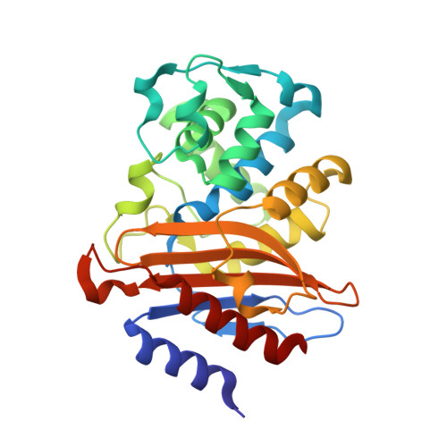
    Evolution leads to conservation of amino acid residues in protein families. Conserved proline residues are usually considered to ensure the correct folding and to stabilize the three-dimensional structure. Surprisingly, proline residues that are highly conserved in class A β-lactamases were found to tolerate various substitutions without large losses in enzyme activity. We investigated the roles of three conserved prolines at positions 107, 226, and 258 in the β-lactamase BlaC from Mycobacterium tuberculosis and found that mutations can lead to dimerization of the enzyme and an overall less stable protein that is prone to aggregate over time. For the variant Pro107Thr, the crystal structure shows dimer formation resembling domain swapping. It is concluded that the proline substitutions loosen the structure, enhancing multimerization. Even though the enzyme does not lose its properties without the conserved proline residues, the prolines ensure the long-term structural integrity of the enzyme.


  • Organizational Affiliation

    Leiden Institute of Chemistry, Leiden University, Leiden, The Netherlands.


Macromolecules
Find similar proteins by:  (by identity cutoff)  |  3D Structure
Entity ID: 1
MoleculeChains Sequence LengthOrganismDetailsImage
Beta-lactamase266Mycobacterium tuberculosisMutation(s): 1 
Gene Names: blaCERS027646_02769
EC: 3.5.2.6
UniProt
Find proteins for P9WKD3 (Mycobacterium tuberculosis (strain ATCC 25618 / H37Rv))
Explore P9WKD3 
Go to UniProtKB:  P9WKD3
Entity Groups  
Sequence Clusters30% Identity50% Identity70% Identity90% Identity95% Identity100% Identity
UniProt GroupP9WKD3
Sequence Annotations
Expand
  • Reference Sequence
Small Molecules
Ligands 3 Unique
IDChains Name / Formula / InChI Key2D Diagram3D Interactions
PO4
Query on PO4

Download Ideal Coordinates CCD File 
K [auth A],
L [auth A]
PHOSPHATE ION
O4 P
NBIIXXVUZAFLBC-UHFFFAOYSA-K
GOL
Query on GOL

Download Ideal Coordinates CCD File 
B [auth A]
C [auth A]
D [auth A]
E [auth A]
F [auth A]
B [auth A],
C [auth A],
D [auth A],
E [auth A],
F [auth A],
G [auth A],
H [auth A],
I [auth A]
GLYCEROL
C3 H8 O3
PEDCQBHIVMGVHV-UHFFFAOYSA-N
EDO
Query on EDO

Download Ideal Coordinates CCD File 
J [auth A]1,2-ETHANEDIOL
C2 H6 O2
LYCAIKOWRPUZTN-UHFFFAOYSA-N
Experimental Data & Validation

Experimental Data

  • Method: X-RAY DIFFRACTION
  • Resolution: 1.30 Å
  • R-Value Free: 0.137 
  • R-Value Work: 0.116 
  • R-Value Observed: 0.117 
  • Space Group: P 21 21 21
Unit Cell:
Length ( Å )Angle ( ˚ )
a = 53.52α = 90
b = 54.231β = 90
c = 79.052γ = 90
Software Package:
Software NamePurpose
REFMACrefinement
XDSdata reduction
Aimlessdata scaling
MOLREPphasing

Structure Validation

View Full Validation Report


Entry History & Funding Information

Deposition Data


Funding OrganizationLocationGrant Number
Netherlands Organisation for Scientific Research (NWO)NetherlandsECHO-711.016.002
H2020 Marie Curie Actions of the European CommissionEuropean Union653706

Revision History  (Full details and data files)

  • Version 1.0: 2021-10-06
    Type: Initial release
  • Version 1.1: 2024-01-31
    Changes: Data collection, Refinement description
  • Version 1.2: 2024-03-27
    Changes: Database references
  • Version 1.3: 2024-04-10
    Changes: Author supporting evidence, Database references