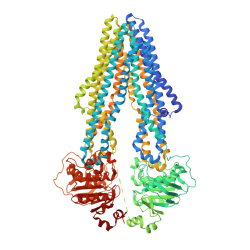
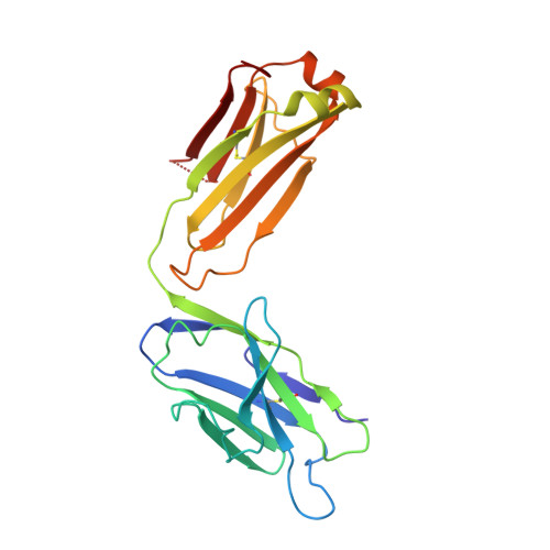
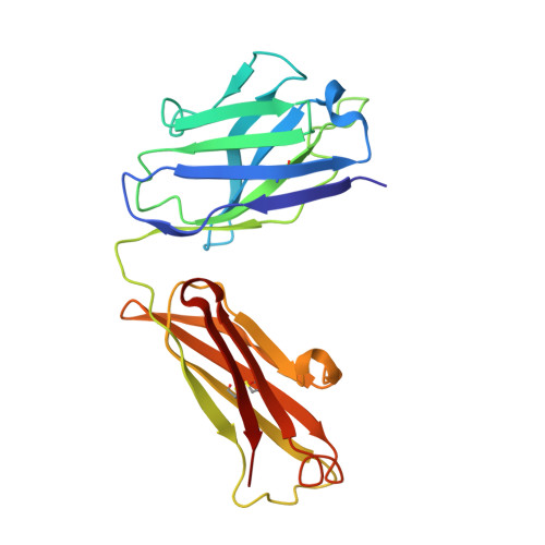
7A6E

Nanodisc reconstituted human ABCB1 in complex with MRK16 Fab and tariquidar


Experimental Data Snapshot

  • Method: ELECTRON MICROSCOPY
  • Resolution: 3.60 Å
  • Aggregation State: PARTICLE 
  • Reconstruction Method: SINGLE PARTICLE 

wwPDB Validation   3D Report Full Report


This is version 1.1 of the entry. See complete history


Literature

Cryo-EM structures reveal distinct mechanisms of inhibition of the human multidrug transporter ABCB1.

Nosol, K.Romane, K.Irobalieva, R.N.Alam, A.Kowal, J.Fujita, N.Locher, K.P.

(2020) Proc Natl Acad Sci U S A 117: 26245-26253

  • DOI: https://doi.org/10.1073/pnas.2010264117
  • Primary Citation of Related Structures:  
    7A65, 7A69, 7A6C, 7A6E, 7A6F

  • PubMed Abstract: 

    ABCB1 detoxifies cells by exporting diverse xenobiotic compounds, thereby limiting drug disposition and contributing to multidrug resistance in cancer cells. Multiple small-molecule inhibitors and inhibitory antibodies have been developed for therapeutic applications, but the structural basis of their activity is insufficiently understood. We determined cryo-EM structures of nanodisc-reconstituted, human ABCB1 in complex with the Fab fragment of the inhibitory, monoclonal antibody MRK16 and bound to a substrate (the antitumor drug vincristine) or to the potent inhibitors elacridar, tariquidar, or zosuquidar. We found that inhibitors bound in pairs, with one molecule lodged in the central drug-binding pocket and a second extending into a phenylalanine-rich cavity that we termed the "access tunnel." This finding explains how inhibitors can act as substrates at low concentration, but interfere with the early steps of the peristaltic extrusion mechanism at higher concentration. Our structural data will also help the development of more potent and selective ABCB1 inhibitors.


  • Organizational Affiliation

    Institute of Molecular Biology and Biophysics, ETH Zürich, 8093 Zürich, Switzerland.


Macromolecules
Find similar proteins by:  (by identity cutoff)  |  3D Structure
Entity ID: 1
MoleculeChains Sequence LengthOrganismDetailsImage
Multidrug resistance protein 11,280Homo sapiensMutation(s): 1 
Gene Names: ABCB1MDR1PGY1
EC: 7.6.2.2 (PDB Primary Data), 7.6.2.1 (PDB Primary Data)
Membrane Entity: Yes 
UniProt & NIH Common Fund Data Resources
Find proteins for P08183 (Homo sapiens)
Explore P08183 
Go to UniProtKB:  P08183
PHAROS:  P08183
GTEx:  ENSG00000085563 
Entity Groups  
Sequence Clusters30% Identity50% Identity70% Identity90% Identity95% Identity100% Identity
UniProt GroupP08183
Sequence Annotations
Expand
  • Reference Sequence
Find similar proteins by:  (by identity cutoff)  |  3D Structure
Entity ID: 2
MoleculeChains Sequence LengthOrganismDetailsImage
MRK16 Fab-fragment light chain219Mus musculusMutation(s): 0 
Entity Groups  
Sequence Clusters30% Identity50% Identity70% Identity90% Identity95% Identity100% Identity
Sequence Annotations
Expand
  • Reference Sequence
Find similar proteins by:  (by identity cutoff)  |  3D Structure
Entity ID: 3
MoleculeChains Sequence LengthOrganismDetailsImage
MRK16 Fab-fragment heavy chain218Mus musculusMutation(s): 0 
Entity Groups  
Sequence Clusters30% Identity50% Identity70% Identity90% Identity95% Identity100% Identity
Sequence Annotations
Expand
  • Reference Sequence
Small Molecules
Ligands 2 Unique
IDChains Name / Formula / InChI Key2D Diagram3D Interactions
R1H (Subject of Investigation/LOI)
Query on R1H

Download Ideal Coordinates CCD File 
D [auth A],
E [auth A]
tariquidar
C38 H38 N4 O6
LGGHDPFKSSRQNS-UHFFFAOYSA-N
CLR
Query on CLR

Download Ideal Coordinates CCD File 
F [auth A]
G [auth A]
H [auth A]
I [auth A]
J [auth A]
F [auth A],
G [auth A],
H [auth A],
I [auth A],
J [auth A],
K [auth A],
L [auth A],
M [auth A],
N [auth A],
O [auth A],
P [auth A]
CHOLESTEROL
C27 H46 O
HVYWMOMLDIMFJA-DPAQBDIFSA-N
Binding Affinity Annotations 
IDSourceBinding Affinity
R1H BindingDB:  7A6E IC50: min: 0.22, max: 1970 (nM) from 24 assay(s)
EC50: min: 16, max: 211 (nM) from 4 assay(s)
CLR BindingDB:  7A6E IC50: 8200 (nM) from 1 assay(s)
Experimental Data & Validation

Experimental Data

  • Method: ELECTRON MICROSCOPY
  • Resolution: 3.60 Å
  • Aggregation State: PARTICLE 
  • Reconstruction Method: SINGLE PARTICLE 

Structure Validation

View Full Validation Report


Entry History & Funding Information

Deposition Data


Funding OrganizationLocationGrant Number
Swiss National Science FoundationSwitzerland310030_189111

Revision History  (Full details and data files)

  • Version 1.0: 2020-10-14
    Type: Initial release
  • Version 1.1: 2020-10-28
    Changes: Database references