7A5W

Structure of D172N BlaC from Mycobacterium tuberculosis


Experimental Data Snapshot

  • Method: X-RAY DIFFRACTION
  • Resolution: 1.40 Å
  • R-Value Free: 0.172 
  • R-Value Work: 0.135 
  • R-Value Observed: 0.136 

wwPDB Validation   3D Report Full Report


This is version 1.3 of the entry. See complete history


Literature

The G132S Mutation Enhances the Resistance of Mycobacterium tuberculosis beta-Lactamase against Sulbactam.

van Alen, I.Chikunova, A.Safeer, A.A.Ahmad, M.U.D.Perrakis, A.Ubbink, M.

(2021) Biochemistry 60: 2236-2245

  • DOI: https://doi.org/10.1021/acs.biochem.1c00168
  • Primary Citation of Related Structures:  
    7A5T, 7A5W, 7A71, 7A72

  • PubMed Abstract: 

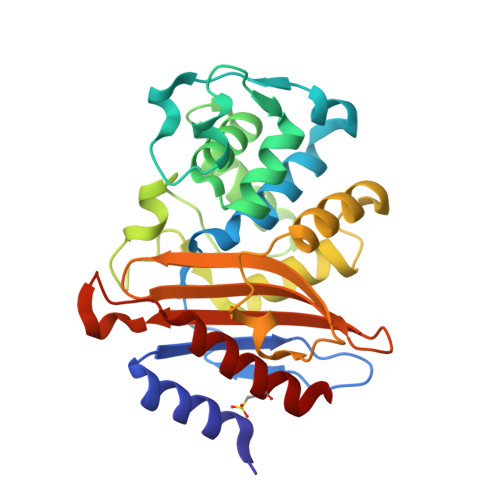
    The current rise of antibiotic resistant forms of Mycobacterium tuberculosis is a global health threat that calls for new antibiotics. The β-lactamase BlaC of this pathogen prevents the use of β-lactam antibiotics, except in combination with a β-lactamase inhibitor. To understand if exposure to such inhibitors can easily result in resistance, a BlaC evolution experiment was performed, studying the evolutionary adaptability against the inhibitor sulbactam. Several amino acid substitutions in BlaC were shown to confer reduced sensitivity to sulbactam. The G132S mutation causes a reduction in the rate of nitrocefin and ampicillin hydrolysis and simultaneously reduces the sensitivity for sulbactam inhibition. Introduction of the side chain moiety of Ser132 causes the 104-105 peptide bond to assume the cis conformation and the side chain of Ser104 to be rotated toward the sulbactam adduct with which it forms a hydrogen bond not present in the wild-type enzyme. The gatekeeper residue Ile105 also moves. These changes in the entrance of the active site can explain the decreased affinity of G132S BlaC for both substrates and sulbactam. Our results show that BlaC can easily acquire a reduced sensitivity for sulbactam, with a single-amino acid mutation, which could hinder the use of combination therapies.


  • Organizational Affiliation

    Leiden Institute of Chemistry, Leiden University, Einsteinweg 55, 2333CC Leiden, The Netherlands.


Macromolecules
Find similar proteins by:  (by identity cutoff)  |  3D Structure
Entity ID: 1
MoleculeChains Sequence LengthOrganismDetailsImage
Beta-lactamase266Mycobacterium tuberculosisMutation(s): 1 
Gene Names: blaCERS027646_02769
EC: 3.5.2.6
UniProt
Find proteins for P9WKD3 (Mycobacterium tuberculosis (strain ATCC 25618 / H37Rv))
Explore P9WKD3 
Go to UniProtKB:  P9WKD3
Entity Groups  
Sequence Clusters30% Identity50% Identity70% Identity90% Identity95% Identity100% Identity
UniProt GroupP9WKD3
Sequence Annotations
Expand
  • Reference Sequence
Experimental Data & Validation

Experimental Data

  • Method: X-RAY DIFFRACTION
  • Resolution: 1.40 Å
  • R-Value Free: 0.172 
  • R-Value Work: 0.135 
  • R-Value Observed: 0.136 
  • Space Group: P 21 21 21
Unit Cell:
Length ( Å )Angle ( ˚ )
a = 54.028α = 90
b = 54.469β = 90
c = 78.78γ = 90
Software Package:
Software NamePurpose
REFMACrefinement
XDSdata reduction
Aimlessdata scaling
MOLREPphasing

Structure Validation

View Full Validation Report


Entry History & Funding Information

Deposition Data


Funding OrganizationLocationGrant Number
Netherlands Organisation for Scientific Research (NWO)NetherlandsECHO-711.016.002
H2020 Marie Curie Actions of the European CommissionNetherlands653706

Revision History  (Full details and data files)

  • Version 1.0: 2021-07-07
    Type: Initial release
  • Version 1.1: 2021-07-21
    Changes: Database references
  • Version 1.2: 2021-07-28
    Changes: Database references
  • Version 1.3: 2024-01-31
    Changes: Data collection, Database references, Refinement description