6ZVN

Botulinum neurotoxin B2 binding domain in complex with human synaptotagmin I


Experimental Data Snapshot

  • Method: X-RAY DIFFRACTION
  • Resolution: 2.50 Å
  • R-Value Free: 0.283 
  • R-Value Work: 0.259 

wwPDB Validation   3D Report Full Report


This is version 2.1 of the entry. See complete history


Literature

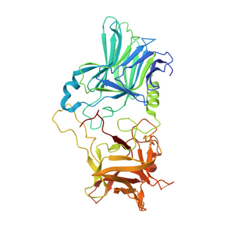
Structural and Biochemical Characterization of Botulinum Neurotoxin Subtype B2 Binding to Its Receptors.

Davies, J.R.Masuyer, G.Stenmark, P.

(2020) Toxins (Basel) 12

  • DOI: https://doi.org/10.3390/toxins12090603
  • Primary Citation of Related Structures:  
    6ZVM, 6ZVN

  • PubMed Abstract: 

    Botulinum neurotoxins (BoNTs) can be used therapeutically to treat a wide range of neuromuscular and neurological conditions. A collection of natural BoNT variants exists which can be classified into serologically distinct serotypes (BoNT/B), and further divided into subtypes (BoNT/B1, B2, …). BoNT subtypes share a high degree of sequence identity within the same serotype yet can display large variation in toxicity. One such example is BoNT/B2, which was isolated from Clostridium botulinum strain 111 in a clinical case of botulism, and presents a 10-fold lower toxicity than BoNT/B1. In an effort to understand the molecular mechanisms behind this difference in potency, we here present the crystal structures of BoNT/B2 in complex with the ganglioside receptor GD1a, and with the human synaptotagmin I protein receptor. We show, using receptor-binding assays, that BoNT/B2 has a slightly higher affinity for GD1a than BoNT/B1, and confirm its considerably weaker affinity for its protein receptors. Although the overall receptor-binding mechanism is conserved for both receptors, structural analysis suggests the lower affinity of BoNT/B2 is the result of key substitutions, where hydrophobic interactions important for synaptotagmin-binding are replaced by polar residues. This study provides a template to drive the development of future BoNT therapeutic molecules centered on assessing the natural subtype variations in receptor-binding that appears to be one of the principal stages driving toxicity.


  • Organizational Affiliation

    Department of Biochemistry and Biophysics, Stockholm University, SE-106 91 Stockholm, Sweden.


Macromolecules
Find similar proteins by:  (by identity cutoff)  |  3D Structure
Entity ID: 1
MoleculeChains Sequence LengthOrganismDetailsImage
NeurotoxinA [auth AAA]438Clostridium botulinumMutation(s): 0 
Gene Names: bontb
UniProt
Find proteins for Q8GR96 (Clostridium botulinum)
Explore Q8GR96 
Go to UniProtKB:  Q8GR96
Entity Groups  
Sequence Clusters30% Identity50% Identity70% Identity90% Identity95% Identity100% Identity
UniProt GroupQ8GR96
Sequence Annotations
Expand
  • Reference Sequence

Find similar proteins by:  Sequence   |   3D Structure  

Entity ID: 2
MoleculeChains Sequence LengthOrganismDetailsImage
Synaptotagmin-1B [auth BBB]21Homo sapiensMutation(s): 0 
UniProt & NIH Common Fund Data Resources
Find proteins for P21579 (Homo sapiens)
Explore P21579 
Go to UniProtKB:  P21579
PHAROS:  P21579
GTEx:  ENSG00000067715 
Entity Groups  
Sequence Clusters30% Identity50% Identity70% Identity90% Identity95% Identity100% Identity
UniProt GroupP21579
Sequence Annotations
Expand
  • Reference Sequence
Experimental Data & Validation

Experimental Data

  • Method: X-RAY DIFFRACTION
  • Resolution: 2.50 Å
  • R-Value Free: 0.283 
  • R-Value Work: 0.259 
  • Space Group: P 21 21 21
Unit Cell:
Length ( Å )Angle ( ˚ )
a = 57.677α = 90
b = 82.528β = 90
c = 106.7γ = 90
Software Package:
Software NamePurpose
REFMACrefinement
DIALSdata reduction
Aimlessdata scaling
PHASERphasing

Structure Validation

View Full Validation Report


Entry History 

Deposition Data

Revision History  (Full details and data files)

  • Version 1.0: 2020-10-14
    Type: Initial release
  • Version 2.0: 2022-09-07
    Changes: Advisory, Atomic model, Database references, Derived calculations, Structure summary
  • Version 2.1: 2024-01-31
    Changes: Data collection, Refinement description