6ZVF

Crystal structure of the recombinant Fab fragment derived from the hybridoma M3/38 in complex with a human Galectin-3 peptide


Experimental Data Snapshot

  • Method: X-RAY DIFFRACTION
  • Resolution: 1.90 Å
  • R-Value Free: 0.243 
  • R-Value Work: 0.203 
  • R-Value Observed: 0.205 

wwPDB Validation   3D Report Full Report


This is version 1.1 of the entry. See complete history


Literature

Effective rational humanization of a PASylated anti-galectin-3 Fab for the sensitive PET imaging of thyroid cancer in vivo.

Peplau, E.De Rose, F.Eichinger, A.Reder, S.Mittelhauser, M.Scafetta, G.Schwaiger, M.Weber, W.A.Bartolazzi, A.D'Alessandria, C.Skerra, A.

(2021) Sci Rep 11: 7358-7358

  • DOI: https://doi.org/10.1038/s41598-021-86641-0
  • Primary Citation of Related Structures:  
    6ZVF

  • PubMed Abstract: 

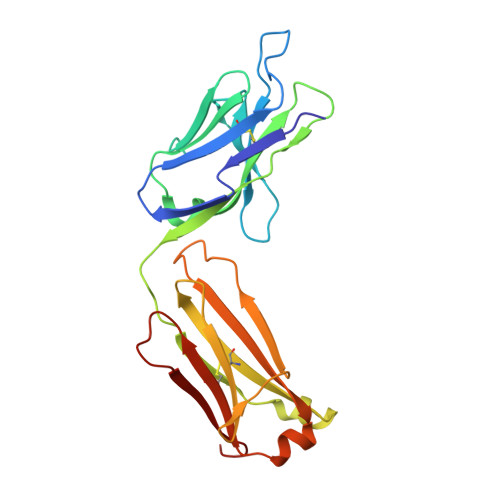
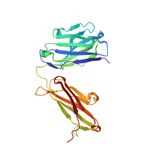
    The lack of a non-invasive test for malignant thyroid nodules makes the diagnosis of thyroid cancer (TC) challenging. Human galectin-3 (hGal3) has emerged as a promising target for medical TC imaging and diagnosis because of its exclusive overexpression in malignant thyroid tissues. We previously developed a human-chimeric αhGal3 Fab fragment derived from the rat monoclonal antibody (mAb) M3/38 with optimized clearance characteristics using PASylation technology. Here, we describe the elucidation of the hGal3 epitope recognized by mAb M3/38, X-ray crystallographic analysis of its complex with the chimeric Fab and, based on the three-dimensional structure, the rational humanization of the Fab by CDR grafting. Four CDR-grafted versions were designed using structurally most closely related fully human immunoglobulin V H /V L regions of which one-employing the acceptor framework regions of the HIV-1 neutralizing human antibody m66-showed the highest antigen affinity. By introducing two additional back-mutations to the rodent donor sequence, an affinity toward hGal3 indistinguishable from the chimeric Fab was achieved (K D  = 0.34 ± 0.02 nM in SPR). The PASylated humanized Fab was site-specifically labelled with the fluorescent dye Cy7 and applied for the immuno-histochemical staining of human tissue sections representative for different TCs. The same protein was conjugated with the metal chelator Dfo, followed by radiolabelling with 89 Zr(IV). The resulting protein tracer allowed the highly sensitive and specific PET/CT imaging of orthotopic tumors in mice, which was confirmed by quantitative analysis of radiotracer accumulation. Thus, the PASylated humanized αhGal3 Fab offers clinical potential for the diagnostic imaging of TC.


  • Organizational Affiliation

    Lehrstuhl für Biologische Chemie, Technische Universität München, 85354, Freising (Weihenstephan), Germany.


Macromolecules
Find similar proteins by:  (by identity cutoff)  |  3D Structure
Entity ID: 1
MoleculeChains Sequence LengthOrganismDetailsImage
Chimeric Fab M3/38 (L,H)A [auth L]219Rattus norvegicusMutation(s): 0 
Entity Groups  
Sequence Clusters30% Identity50% Identity70% Identity90% Identity95% Identity100% Identity
Sequence Annotations
Expand
  • Reference Sequence
Find similar proteins by:  (by identity cutoff)  |  3D Structure
Entity ID: 2
MoleculeChains Sequence LengthOrganismDetailsImage
Chimeric Fab M3/38 (L,H)B [auth H]227Rattus norvegicusMutation(s): 0 
Entity Groups  
Sequence Clusters30% Identity50% Identity70% Identity90% Identity95% Identity100% Identity
Sequence Annotations
Expand
  • Reference Sequence

Find similar proteins by:  Sequence   |   3D Structure  

Entity ID: 3
MoleculeChains Sequence LengthOrganismDetailsImage
Galectin-3C [auth P]10Homo sapiensMutation(s): 1 
UniProt & NIH Common Fund Data Resources
Find proteins for P17931 (Homo sapiens)
Explore P17931 
Go to UniProtKB:  P17931
PHAROS:  P17931
GTEx:  ENSG00000131981 
Entity Groups  
Sequence Clusters30% Identity50% Identity70% Identity90% Identity95% Identity100% Identity
UniProt GroupP17931
Sequence Annotations
Expand
  • Reference Sequence
Experimental Data & Validation

Experimental Data

  • Method: X-RAY DIFFRACTION
  • Resolution: 1.90 Å
  • R-Value Free: 0.243 
  • R-Value Work: 0.203 
  • R-Value Observed: 0.205 
  • Space Group: C 1 2 1
Unit Cell:
Length ( Å )Angle ( ˚ )
a = 94.134α = 90
b = 61.27β = 103.36
c = 80.909γ = 90
Software Package:
Software NamePurpose
REFMACrefinement
MOSFLMdata reduction
SCALAdata scaling
PHASERphasing
PDB_EXTRACTdata extraction

Structure Validation

View Full Validation Report


Entry History & Funding Information

Deposition Data


Funding OrganizationLocationGrant Number
German Research Foundation (DFG)GermanySK 33/11-1

Revision History  (Full details and data files)

  • Version 1.0: 2021-04-14
    Type: Initial release
  • Version 1.1: 2024-01-31
    Changes: Data collection, Database references, Refinement description