6ZPH

Kinesin binding protein complexed with Kif15 motor domain


Experimental Data Snapshot

  • Method: ELECTRON MICROSCOPY
  • Resolution: 6.90 Å
  • Aggregation State: PARTICLE 
  • Reconstruction Method: SINGLE PARTICLE 

wwPDB Validation   3D Report Full Report


This is version 1.0 of the entry. See complete history


Literature

The mechanism of kinesin inhibition by kinesin-binding protein.

Atherton, J.Hummel, J.J.Olieric, N.Locke, J.Pena, A.Rosenfeld, S.S.Steinmetz, M.O.Hoogenraad, C.C.Moores, C.A.

(2020) Elife 9

  • DOI: https://doi.org/10.7554/eLife.61481
  • Primary Citation of Related Structures:  
    6ZPG, 6ZPH, 6ZPI

  • PubMed Abstract: 

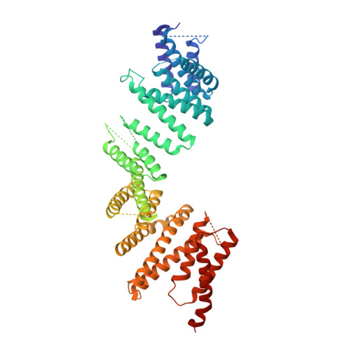
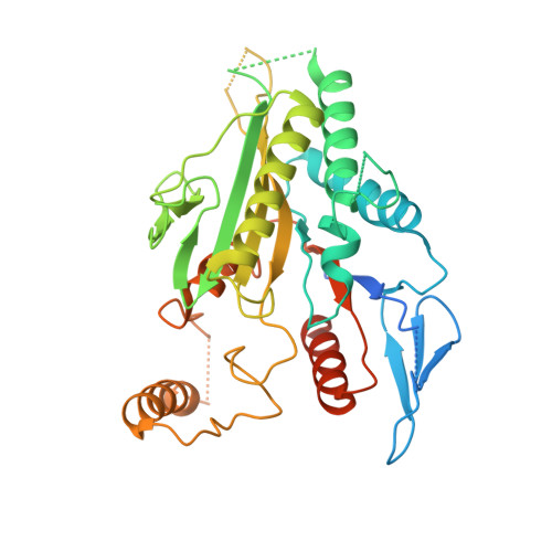
    Subcellular compartmentalisation is necessary for eukaryotic cell function. Spatial and temporal regulation of kinesin activity is essential for building these local environments via control of intracellular cargo distribution. Kinesin-binding protein (KBP) interacts with a subset of kinesins via their motor domains, inhibits their microtubule (MT) attachment, and blocks their cellular function. However, its mechanisms of inhibition and selectivity have been unclear. Here we use cryo-electron microscopy to reveal the structure of KBP and of a KBP-kinesin motor domain complex. KBP is a tetratricopeptide repeat-containing, right-handed α-solenoid that sequesters the kinesin motor domain's tubulin-binding surface, structurally distorting the motor domain and sterically blocking its MT attachment. KBP uses its α-solenoid concave face and edge loops to bind the kinesin motor domain, and selected structure-guided mutations disrupt KBP inhibition of kinesin transport in cells. The KBP-interacting motor domain surface contains motifs exclusively conserved in KBP-interacting kinesins, suggesting a basis for kinesin selectivity.


  • Organizational Affiliation

    Randall Centre for Cell and Molecular Biophysics, King's College, London, United Kingdom.


Macromolecules
Find similar proteins by:  (by identity cutoff)  |  3D Structure
Entity ID: 1
MoleculeChains Sequence LengthOrganismDetailsImage
KIF-binding protein621Homo sapiensMutation(s): 0 
Gene Names: KIFBPKBPKIAA1279KIF1BP
UniProt & NIH Common Fund Data Resources
Find proteins for Q96EK5 (Homo sapiens)
Explore Q96EK5 
Go to UniProtKB:  Q96EK5
PHAROS:  Q96EK5
GTEx:  ENSG00000198954 
Entity Groups  
Sequence Clusters30% Identity50% Identity70% Identity90% Identity95% Identity100% Identity
UniProt GroupQ96EK5
Sequence Annotations
Expand
  • Reference Sequence
Find similar proteins by:  (by identity cutoff)  |  3D Structure
Entity ID: 2
MoleculeChains Sequence LengthOrganismDetailsImage
Kinesin-like protein KIF15383Homo sapiensMutation(s): 0 
Gene Names: KIF15KLP2KNSL7
UniProt & NIH Common Fund Data Resources
Find proteins for Q9NS87 (Homo sapiens)
Explore Q9NS87 
Go to UniProtKB:  Q9NS87
PHAROS:  Q9NS87
GTEx:  ENSG00000163808 
Entity Groups  
Sequence Clusters30% Identity50% Identity70% Identity90% Identity95% Identity100% Identity
UniProt GroupQ9NS87
Sequence Annotations
Expand
  • Reference Sequence
Small Molecules
Ligands 2 Unique
IDChains Name / Formula / InChI Key2D Diagram3D Interactions
ADP
Query on ADP

Download Ideal Coordinates CCD File 
C [auth B]ADENOSINE-5'-DIPHOSPHATE
C10 H15 N5 O10 P2
XTWYTFMLZFPYCI-KQYNXXCUSA-N
MG
Query on MG

Download Ideal Coordinates CCD File 
D [auth B]MAGNESIUM ION
Mg
JLVVSXFLKOJNIY-UHFFFAOYSA-N
Experimental Data & Validation

Experimental Data

  • Method: ELECTRON MICROSCOPY
  • Resolution: 6.90 Å
  • Aggregation State: PARTICLE 
  • Reconstruction Method: SINGLE PARTICLE 

Structure Validation

View Full Validation Report


Entry History & Funding Information

Deposition Data


Funding OrganizationLocationGrant Number
Medical Research Council (MRC, United Kingdom)United KingdomMR/R000352/1
Worldwide Cancer ResearchUnited Kingdom16-0037
Swiss National Science FoundationSwitzerland31003A_166608
Wellcome TrustUnited Kingdom202679/Z/16/Z
Wellcome TrustUnited Kingdom206166/Z/17/Z
National Institutes of Health/National Institute of General Medical Sciences (NIH/NIGMS)United StatesR01GM130556

Revision History  (Full details and data files)

  • Version 1.0: 2020-12-30
    Type: Initial release