6ZNA

Porcine ATP synthase Fo domain


Experimental Data Snapshot

  • Method: ELECTRON MICROSCOPY
  • Resolution: 6.20 Å
  • Aggregation State: PARTICLE 
  • Reconstruction Method: SINGLE PARTICLE 

wwPDB Validation   3D Report Full Report


This is version 1.2 of the entry. See complete history


Literature

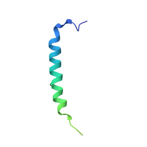
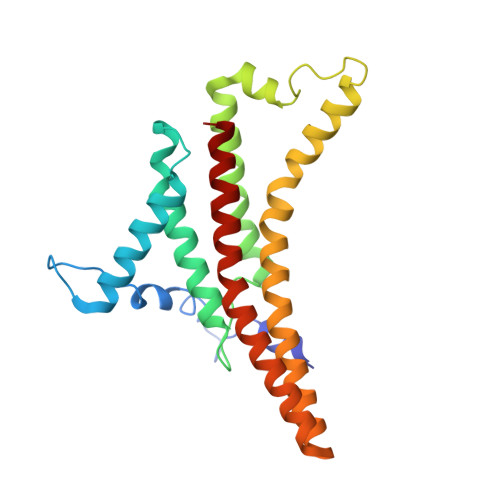
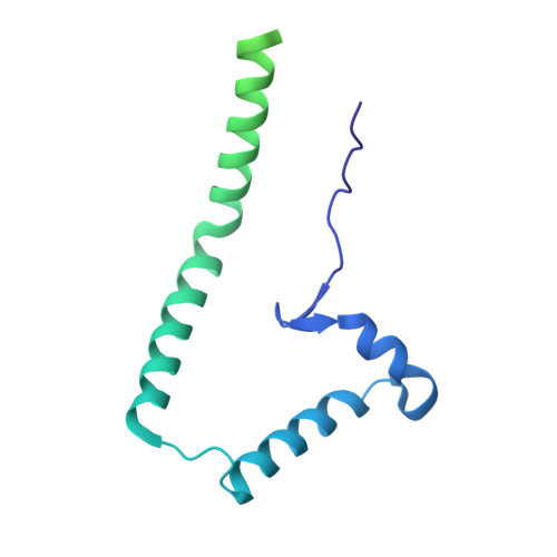
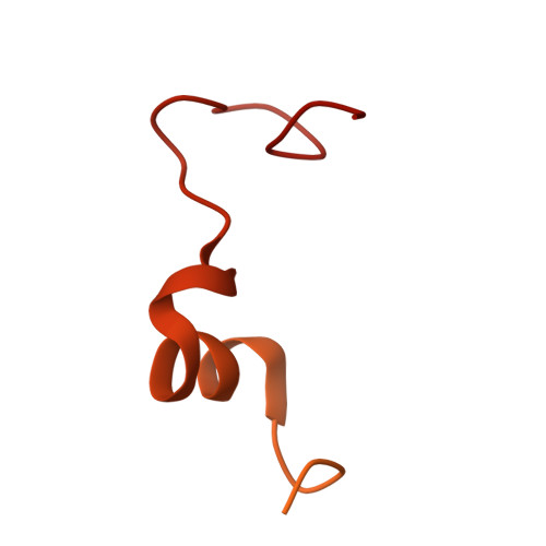
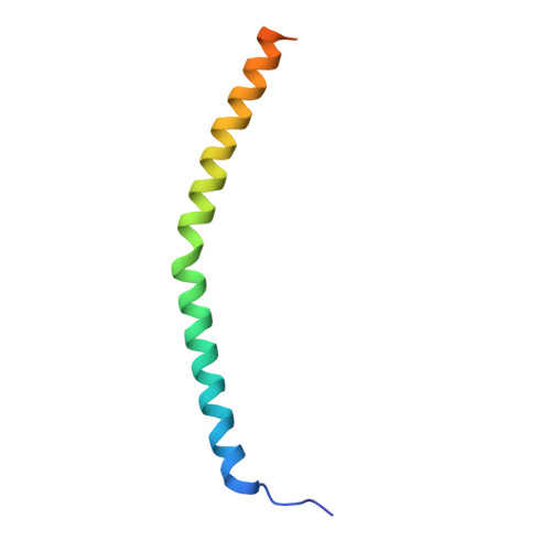
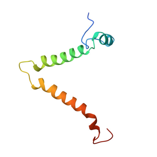
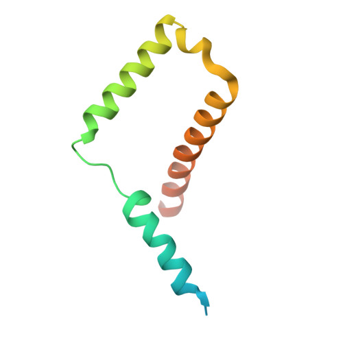
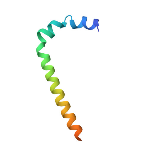
Structure of the dimeric ATP synthase from bovine mitochondria.

Spikes, T.E.Montgomery, M.G.Walker, J.E.

(2020) Proc Natl Acad Sci U S A 117: 23519-23526

  • DOI: https://doi.org/10.1073/pnas.2013998117
  • Primary Citation of Related Structures:  
    6YY0, 6Z1R, 6Z1U, 6ZBB, 6ZG7, 6ZG8, 6ZIK, 6ZIQ, 6ZIT, 6ZIU, 6ZMR, 6ZNA, 6ZPO, 6ZQM, 6ZQN

  • PubMed Abstract: 

    The structure of the dimeric ATP synthase from bovine mitochondria determined in three rotational states by electron cryo-microscopy provides evidence that the proton uptake from the mitochondrial matrix via the proton inlet half channel proceeds via a Grotthus mechanism, and a similar mechanism may operate in the exit half channel. The structure has given information about the architecture and mechanical constitution and properties of the peripheral stalk, part of the membrane extrinsic region of the stator, and how the action of the peripheral stalk damps the side-to-side rocking motions that occur in the enzyme complex during the catalytic cycle. It also describes wedge structures in the membrane domains of each monomer, where the skeleton of each wedge is provided by three α-helices in the membrane domains of the b-subunit to which the supernumerary subunits e, f, and g and the membrane domain of subunit A6L are bound. Protein voids in the wedge are filled by three specifically bound cardiolipin molecules and two other phospholipids. The external surfaces of the wedges link the monomeric complexes together into the dimeric structures and provide a pivot to allow the monomer-monomer interfaces to change during catalysis and to accommodate other changes not related directly to catalysis in the monomer-monomer interface that occur in mitochondrial cristae. The structure of the bovine dimer also demonstrates that the structures of dimeric ATP synthases in a tetrameric porcine enzyme have been seriously misinterpreted in the membrane domains.


  • Organizational Affiliation

    The Medical Research Council Mitochondrial Biology Unit, Cambridge Biomedical Campus, University of Cambridge, Cambridge CB2 0XY, United Kingdom.


Macromolecules
Find similar proteins by:  (by identity cutoff)  |  3D Structure
Entity ID: 1
MoleculeChains Sequence LengthOrganismDetailsImage
ATP synthase protein 8A [auth A8],
IA [auth C8],
NB [auth 8],
R [auth B8]
67Sus scrofaMutation(s): 0 
Membrane Entity: Yes 
UniProt
Find proteins for Q35914 (Sus scrofa)
Explore Q35914 
Go to UniProtKB:  Q35914
Entity Groups  
Sequence Clusters30% Identity50% Identity70% Identity90% Identity95% Identity100% Identity
UniProt GroupQ35914
Sequence Annotations
Expand
  • Reference Sequence
Find similar proteins by:  (by identity cutoff)  |  3D Structure
Entity ID: 2
MoleculeChains Sequence LengthOrganismDetailsImage
ATP synthase F(0) complex subunit C1, mitochondrial75Sus scrofaMutation(s): 0 
Membrane Entity: Yes 
UniProt
Find proteins for A0A8D1N484 (Sus scrofa)
Explore A0A8D1N484 
Go to UniProtKB:  A0A8D1N484
Entity Groups  
Sequence Clusters30% Identity50% Identity70% Identity90% Identity95% Identity100% Identity
UniProt GroupA0A8D1N484
Sequence Annotations
Expand
  • Reference Sequence
Find similar proteins by:  (by identity cutoff)  |  3D Structure
Entity ID: 3
MoleculeChains Sequence LengthOrganismDetailsImage
ATP synthase subunit aAA [auth Ba],
HB [auth a],
J [auth Aa],
RA [auth Ca]
226Sus scrofaMutation(s): 0 
Membrane Entity: Yes 
UniProt
Find proteins for Q35915 (Sus scrofa)
Explore Q35915 
Go to UniProtKB:  Q35915
Entity Groups  
Sequence Clusters30% Identity50% Identity70% Identity90% Identity95% Identity100% Identity
UniProt GroupQ35915
Sequence Annotations
Expand
  • Reference Sequence
Find similar proteins by:  (by identity cutoff)  |  3D Structure
Entity ID: 4
MoleculeChains Sequence LengthOrganismDetailsImage
ATP synthase peripheral stalk-membrane subunit bBA [auth Bb],
IB [auth b],
K [auth Ab],
SA [auth Cb]
214Sus scrofaMutation(s): 0 
Membrane Entity: Yes 
UniProt
Find proteins for A0A8D0MI69 (Sus scrofa)
Explore A0A8D0MI69 
Go to UniProtKB:  A0A8D0MI69
Entity Groups  
Sequence Clusters30% Identity50% Identity70% Identity90% Identity95% Identity100% Identity
UniProt GroupA0A8D0MI69
Sequence Annotations
Expand
  • Reference Sequence
Find similar proteins by:  (by identity cutoff)  |  3D Structure
Entity ID: 5
MoleculeChains Sequence LengthOrganismDetailsImage
ATP synthase subunit d, mitochondrialCA [auth Bd],
JB [auth d],
L [auth Ad],
TA [auth Cd]
160Sus scrofaMutation(s): 0 
Membrane Entity: Yes 
UniProt
Find proteins for A0A4X1V3L8 (Sus scrofa)
Explore A0A4X1V3L8 
Go to UniProtKB:  A0A4X1V3L8
Entity Groups  
Sequence Clusters30% Identity50% Identity70% Identity90% Identity95% Identity100% Identity
UniProt GroupA0A4X1V3L8
Sequence Annotations
Expand
  • Reference Sequence
Find similar proteins by:  (by identity cutoff)  |  3D Structure
Entity ID: 6
MoleculeChains Sequence LengthOrganismDetailsImage
ATP synthase subunit e, mitochondrialDA [auth Be],
KB [auth e],
M [auth Ae],
UA [auth Ce]
70Sus scrofaMutation(s): 0 
Membrane Entity: Yes 
UniProt
Find proteins for Q9MYT8 (Sus scrofa)
Explore Q9MYT8 
Go to UniProtKB:  Q9MYT8
Entity Groups  
Sequence Clusters30% Identity50% Identity70% Identity90% Identity95% Identity100% Identity
UniProt GroupQ9MYT8
Sequence Annotations
Expand
  • Reference Sequence
Find similar proteins by:  (by identity cutoff)  |  3D Structure
Entity ID: 7
MoleculeChains Sequence LengthOrganismDetailsImage
ATP synthase subunit f, mitochondrialEA [auth Bf],
LB [auth f],
N [auth Af],
VA [auth Cf]
87Sus scrofaMutation(s): 0 
Membrane Entity: Yes 
UniProt
Find proteins for Q95339 (Sus scrofa)
Explore Q95339 
Go to UniProtKB:  Q95339
Entity Groups  
Sequence Clusters30% Identity50% Identity70% Identity90% Identity95% Identity100% Identity
UniProt GroupQ95339
Sequence Annotations
Expand
  • Reference Sequence
Find similar proteins by:  (by identity cutoff)  |  3D Structure
Entity ID: 8
MoleculeChains Sequence LengthOrganismDetailsImage
ATP synthase g subunitFA [auth Bg],
MB [auth g],
O [auth Ag],
WA [auth Cg]
102Sus scrofaMutation(s): 0 
Membrane Entity: Yes 
UniProt
Find proteins for F1SAK7 (Sus scrofa)
Explore F1SAK7 
Go to UniProtKB:  F1SAK7
Entity Groups  
Sequence Clusters30% Identity50% Identity70% Identity90% Identity95% Identity100% Identity
UniProt GroupF1SAK7
Sequence Annotations
Expand
  • Reference Sequence
Find similar proteins by:  (by identity cutoff)  |  3D Structure
Entity ID: 9
MoleculeChains Sequence LengthOrganismDetailsImage
ATP synthase j subunit (6.8PL)GA [auth Bj],
OB [auth j],
P [auth Aj],
XA [auth Cj]
60Sus scrofaMutation(s): 0 
Membrane Entity: Yes 
UniProt
Find proteins for A0A4X1TX70 (Sus scrofa)
Explore A0A4X1TX70 
Go to UniProtKB:  A0A4X1TX70
Entity Groups  
Sequence Clusters30% Identity50% Identity70% Identity90% Identity95% Identity100% Identity
UniProt GroupA0A4X1TX70
Sequence Annotations
Expand
  • Reference Sequence
Find similar proteins by:  (by identity cutoff)  |  3D Structure
Entity ID: 10
MoleculeChains Sequence LengthOrganismDetailsImage
ATP synthase membrane subunit DAPITHA [auth Bk],
PB [auth k],
Q [auth Ak],
YA [auth Ck]
57Sus scrofaMutation(s): 0 
Membrane Entity: Yes 
UniProt
Find proteins for A0A4X1TQQ5 (Sus scrofa)
Explore A0A4X1TQQ5 
Go to UniProtKB:  A0A4X1TQQ5
Entity Groups  
Sequence Clusters30% Identity50% Identity70% Identity90% Identity95% Identity100% Identity
UniProt GroupA0A4X1TQQ5
Sequence Annotations
Expand
  • Reference Sequence
Small Molecules
Modified Residues  1 Unique
IDChains TypeFormula2D DiagramParent
M3L
Query on M3L
AB [auth L]
B [auth AK]
BB [auth M]
C [auth AL]
CB [auth N]
AB [auth L],
B [auth AK],
BB [auth M],
C [auth AL],
CB [auth N],
D [auth AM],
DB [auth O],
E [auth AN],
EB [auth P],
F [auth AO],
FB [auth Q],
G [auth AP],
GB [auth R],
H [auth AQ],
I [auth AR],
JA [auth CK],
KA [auth CL],
LA [auth CM],
MA [auth CN],
NA [auth CO],
OA [auth CP],
PA [auth CQ],
QA [auth CR],
S [auth BK],
T [auth BL],
U [auth BM],
V [auth BN],
W [auth BO],
X [auth BP],
Y [auth BQ],
Z [auth BR],
ZA [auth K]
L-PEPTIDE LINKINGC9 H21 N2 O2LYS
Experimental Data & Validation

Experimental Data

  • Method: ELECTRON MICROSCOPY
  • Resolution: 6.20 Å
  • Aggregation State: PARTICLE 
  • Reconstruction Method: SINGLE PARTICLE 

Structure Validation

View Full Validation Report


Entry History & Funding Information

Deposition Data


Funding OrganizationLocationGrant Number
Medical Research Council (MRC, United Kingdom)United KingdomMR/M009858/1

Revision History  (Full details and data files)

  • Version 1.0: 2020-09-09
    Type: Initial release
  • Version 1.1: 2020-09-23
    Changes: Database references
  • Version 1.2: 2020-09-30
    Changes: Database references