6ZMZ

UDPG-bound Trehalose transferase from Thermoproteus uzoniensis


Experimental Data Snapshot

  • Method: X-RAY DIFFRACTION
  • Resolution: 1.90 Å
  • R-Value Free: 0.223 
  • R-Value Work: 0.185 

wwPDB Validation   3D Report Full Report


Ligand Structure Quality Assessment 


This is version 1.0 of the entry. See complete history


Literature

Anomeric Selectivity of Trehalose Transferase with Rare l-Sugars.

Mestrom, L.Marsden, S.R.van der Eijk, H.Laustsen, J.U.Jeffries, C.M.Svergun, D.I.Hagedoorn, P.L.Bento, I.Hanefeld, U.

(2020) ACS Catal 10: 8835-8839

  • DOI: https://doi.org/10.1021/acscatal.0c02117
  • Primary Citation of Related Structures:  
    6ZJ4, 6ZJ7, 6ZMZ

  • PubMed Abstract: 

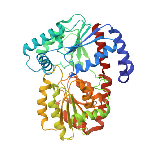
    Retaining LeLoir glycosyltransferases catalyze the formation of glycosidic bonds between nucleotide sugar donors and carbohydrate acceptors. The anomeric selectivity of trehalose transferase from Thermoproteus uzoniensis was investigated for both d- and l-glycopyranose acceptors. The enzyme couples a wide range of carbohydrates, yielding trehalose analogues with conversion and enantioselectivity of >98%. The anomeric selectivity inverts from α,α-(1 → 1)-glycosidic bonds for d-glycopyranose acceptors to α,β-(1 → 1)-glycosidic bonds for l-glycopyranose acceptors, while ( S )-selectivity was retained for both types of sugar acceptors. Comparison of protein crystal structures of trehalose transferase in complex with α,α-trehalose and an unnatural α,β-trehalose analogue highlighted the mechanistic rationale for the observed inversion of anomeric selectivity.


  • Organizational Affiliation

    Department of Biotechnology, Delft University of Technology, Van der Maasweg 9, 2629 HZ, Delft, The Netherlands.


Macromolecules
Find similar proteins by:  (by identity cutoff)  |  3D Structure
Entity ID: 1
MoleculeChains Sequence LengthOrganismDetailsImage
Trehalose phosphorylase/synthaseA [auth AAA]400Thermoproteus uzoniensisMutation(s): 0 
Gene Names: TUZN_0976
UniProt
Find proteins for F2L613 (Thermoproteus uzoniensis (strain 768-20))
Explore F2L613 
Go to UniProtKB:  F2L613
Entity Groups  
Sequence Clusters30% Identity50% Identity70% Identity90% Identity95% Identity100% Identity
UniProt GroupF2L613
Sequence Annotations
Expand
  • Reference Sequence
Small Molecules
Ligands 2 Unique
IDChains Name / Formula / InChI Key2D Diagram3D Interactions
UPG (Subject of Investigation/LOI)
Query on UPG

Download Ideal Coordinates CCD File 
B [auth AAA]URIDINE-5'-DIPHOSPHATE-GLUCOSE
C15 H24 N2 O17 P2
HSCJRCZFDFQWRP-JZMIEXBBSA-N
PEG
Query on PEG

Download Ideal Coordinates CCD File 
C [auth AAA]DI(HYDROXYETHYL)ETHER
C4 H10 O3
MTHSVFCYNBDYFN-UHFFFAOYSA-N
Experimental Data & Validation

Experimental Data

  • Method: X-RAY DIFFRACTION
  • Resolution: 1.90 Å
  • R-Value Free: 0.223 
  • R-Value Work: 0.185 
  • Space Group: P 21 21 21
Unit Cell:
Length ( Å )Angle ( ˚ )
a = 53.045α = 90
b = 69.327β = 90
c = 98.912γ = 90
Software Package:
Software NamePurpose
REFMACrefinement
XDSdata reduction
Aimlessdata scaling
MOLREPphasing

Structure Validation

View Full Validation Report


Ligand Structure Quality Assessment 


Entry History & Funding Information

Deposition Data


Funding OrganizationLocationGrant Number
European CommissionGermanyERA-IB-15-110

Revision History  (Full details and data files)

  • Version 1.0: 2020-09-30
    Type: Initial release