6ZLL

Crystal Structure of UDP-Glucuronic acid 4-epimerase from Bacillus cereus in complex with UDP-Galacturonic acid and NAD


Experimental Data Snapshot

  • Method: X-RAY DIFFRACTION
  • Resolution: 1.85 Å
  • R-Value Free: 0.222 
  • R-Value Work: 0.187 
  • R-Value Observed: 0.189 

wwPDB Validation   3D Report Full Report


Ligand Structure Quality Assessment 


This is version 1.2 of the entry. See complete history


Literature

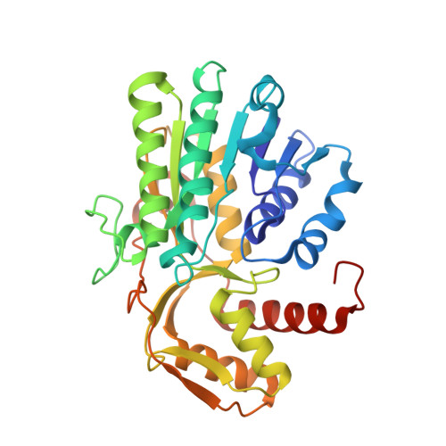
Crystallographic snapshots of UDP-glucuronic acid 4-epimerase ligand binding, rotation, and reduction.

Iacovino, L.G.Savino, S.Borg, A.J.E.Binda, C.Nidetzky, B.Mattevi, A.

(2020) J Biol Chem 295: 12461-12473

  • DOI: https://doi.org/10.1074/jbc.RA120.014692
  • Primary Citation of Related Structures:  
    6ZL6, 6ZLA, 6ZLD, 6ZLJ, 6ZLK, 6ZLL

  • PubMed Abstract: 

    UDP-glucuronic acid is converted to UDP-galacturonic acid en route to a variety of sugar-containing metabolites. This reaction is performed by a NAD + -dependent epimerase belonging to the short-chain dehydrogenase/reductase family. We present several high-resolution crystal structures of the UDP-glucuronic acid epimerase from Bacillus cereus The geometry of the substrate-NAD + interactions is finely arranged to promote hydride transfer. The exquisite complementarity between glucuronic acid and its binding site is highlighted by the observation that the unligated cavity is occupied by a cluster of ordered waters whose positions overlap the polar groups of the sugar substrate. Co-crystallization experiments led to a structure where substrate- and product-bound enzymes coexist within the same crystal. This equilibrium structure reveals the basis for a "swing and flip" rotation of the pro-chiral 4-keto-hexose-uronic acid intermediate that results from glucuronic acid oxidation, placing the C4' atom in position for receiving a hydride ion on the opposite side of the sugar ring. The product-bound active site is almost identical to that of the substrate-bound structure and satisfies all hydrogen-bonding requirements of the ligand. The structure of the apoenzyme together with the kinetic isotope effect and mutagenesis experiments further outlines a few flexible loops that exist in discrete conformations, imparting structural malleability required for ligand rotation while avoiding leakage of the catalytic intermediate and/or side reactions. These data highlight the double nature of the enzymatic mechanism: the active site features a high degree of precision in substrate recognition combined with the flexibility required for intermediate rotation.


  • Organizational Affiliation

    Department of Biology and Biotechnology "Lazzaro Spallanzani", University of Pavia, Pavia, Italy.


Macromolecules
Find similar proteins by:  (by identity cutoff)  |  3D Structure
Entity ID: 1
MoleculeChains Sequence LengthOrganismDetailsImage
Epimerase domain-containing protein
A, B, C, D
327Bacillus cereus HuA2-4Mutation(s): 0 
Gene Names: IG7_05634
Entity Groups  
Sequence Clusters30% Identity50% Identity70% Identity90% Identity95% Identity100% Identity
Sequence Annotations
Expand
  • Reference Sequence
Small Molecules
Ligands 2 Unique
IDChains Name / Formula / InChI Key2D Diagram3D Interactions
NAD (Subject of Investigation/LOI)
Query on NAD

Download Ideal Coordinates CCD File 
F [auth A],
H [auth B],
J [auth C],
L [auth D]
NICOTINAMIDE-ADENINE-DINUCLEOTIDE
C21 H27 N7 O14 P2
BAWFJGJZGIEFAR-NNYOXOHSSA-N
UGB (Subject of Investigation/LOI)
Query on UGB

Download Ideal Coordinates CCD File 
E [auth A],
G [auth B],
I [auth C],
K [auth D]
(2S,3R,4S,5R,6R)-6-[[[(2R,3S,4R,5R)-5-(2,4-dioxopyrimidin-1-yl)-3,4-dihydroxy-oxolan-2-yl]methoxy-hydroxy-phosphoryl]oxy-hydroxy-phosphoryl]oxy-3,4,5-trihydroxy-oxane-2-carboxylic acid
C15 H22 N2 O18 P2
HDYANYHVCAPMJV-GXNRKQDOSA-N
Experimental Data & Validation

Experimental Data

  • Method: X-RAY DIFFRACTION
  • Resolution: 1.85 Å
  • R-Value Free: 0.222 
  • R-Value Work: 0.187 
  • R-Value Observed: 0.189 
  • Space Group: P 1
Unit Cell:
Length ( Å )Angle ( ˚ )
a = 56.787α = 91.92
b = 62.652β = 99.86
c = 105.712γ = 91.98
Software Package:
Software NamePurpose
REFMACrefinement
PDB_EXTRACTdata extraction
XDSdata reduction
Aimlessdata scaling
MOLREPphasing

Structure Validation

View Full Validation Report


Ligand Structure Quality Assessment 


Entry History & Funding Information

Deposition Data


Funding OrganizationLocationGrant Number
Italian Ministry of EducationItaly--

Revision History  (Full details and data files)

  • Version 1.0: 2020-07-29
    Type: Initial release
  • Version 1.1: 2020-09-09
    Changes: Database references
  • Version 1.2: 2024-01-31
    Changes: Data collection, Database references, Refinement description