6ZKW

Crystal structure of InhA:01 TCR in complex with HLA-E bound to InhA (53-61)


Experimental Data Snapshot

  • Method: X-RAY DIFFRACTION
  • Resolution: 2.26 Å
  • R-Value Free: 0.287 
  • R-Value Work: 0.243 
  • R-Value Observed: 0.245 

wwPDB Validation   3D Report Full Report


This is version 1.3 of the entry. See complete history


Literature

Structure-guided stabilization of pathogen-derived peptide-HLA-E complexes using non-natural amino acids conserves native TCR recognition.

Barber, C.De Souza, V.A.Paterson, R.L.Martin-Urdiroz, M.Mulakkal, N.C.Srikannathasan, V.Connolly, M.Phillips, G.Foong-Leong, T.Pengelly, R.Karuppiah, V.Grant, T.Dembek, M.Verma, A.Gibbs-Howe, D.Blicher, T.H.Knox, A.Robinson, R.A.Cole, D.K.Leonard, S.

(2022) Eur J Immunol 52: 618-632

  • DOI: https://doi.org/10.1002/eji.202149745
  • Primary Citation of Related Structures:  
    6ZKW, 6ZKX, 6ZKY, 6ZKZ, 7NDQ, 7NDT, 7NDU

  • PubMed Abstract: 

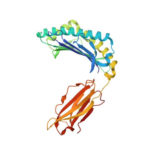
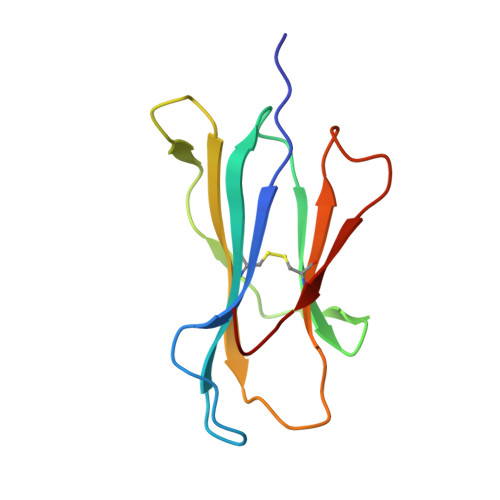
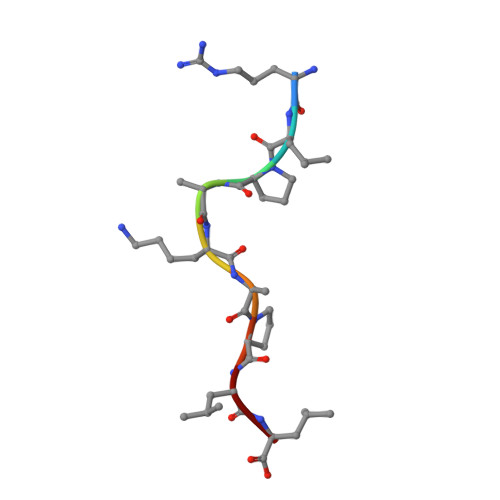
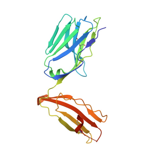
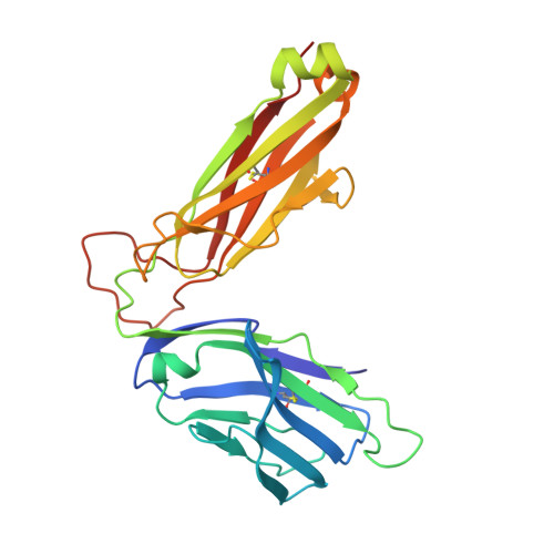
    The nonpolymorphic class Ib molecule, HLA-E, primarily presents peptides from HLA class Ia leader peptides, providing an inhibitory signal to NK cells via CD94/NKG2 interactions. Although peptides of pathogenic origin can also be presented by HLA-E to T cells, the molecular basis underpinning their role in antigen surveillance is largely unknown. Here, we solved a co-complex crystal structure of a TCR with an HLA-E presented peptide (pHLA-E) from bacterial (Mycobacterium tuberculosis) origin, and the first TCR-pHLA-E complex with a noncanonically presented peptide from viral (HIV) origin. The structures provided a molecular foundation to develop a novel method to introduce cysteine traps using non-natural amino acid chemistry that stabilized pHLA-E complexes while maintaining native interface contacts between the TCRs and different pHLA-E complexes. These pHLA-E monomers could be used to isolate pHLA-E-specific T cells, with obvious utility for studying pHLA-E restricted T cells, and for the identification of putative therapeutic TCRs.


  • Organizational Affiliation

    Immunocore Ltd, Abingdon, Oxfordshire, UK.


Macromolecules
Find similar proteins by:  (by identity cutoff)  |  3D Structure
Entity ID: 1
MoleculeChains Sequence LengthOrganismDetailsImage
HLA class I histocompatibility antigen, alpha chain E276Homo sapiensMutation(s): 0 
Gene Names: HLA-EHLA-6.2HLAE
UniProt & NIH Common Fund Data Resources
Find proteins for P13747 (Homo sapiens)
Explore P13747 
Go to UniProtKB:  P13747
PHAROS:  P13747
GTEx:  ENSG00000204592 
Entity Groups  
Sequence Clusters30% Identity50% Identity70% Identity90% Identity95% Identity100% Identity
UniProt GroupP13747
Sequence Annotations
Expand
  • Reference Sequence
Find similar proteins by:  (by identity cutoff)  |  3D Structure
Entity ID: 2
MoleculeChains Sequence LengthOrganismDetailsImage
Beta-2-microglobulin100Homo sapiensMutation(s): 0 
Gene Names: B2MCDABP0092HDCMA22P
UniProt & NIH Common Fund Data Resources
Find proteins for P61769 (Homo sapiens)
Explore P61769 
Go to UniProtKB:  P61769
PHAROS:  P61769
GTEx:  ENSG00000166710 
Entity Groups  
Sequence Clusters30% Identity50% Identity70% Identity90% Identity95% Identity100% Identity
UniProt GroupP61769
Sequence Annotations
Expand
  • Reference Sequence

Find similar proteins by:  Sequence   |   3D Structure  

Entity ID: 3
MoleculeChains Sequence LengthOrganismDetailsImage
Enoyl-[acyl-carrier-protein] reductase [NADH]9Mycobacterium tuberculosis H37RvMutation(s): 0 
EC: 1.3.1.9
UniProt
Find proteins for P9WGR1 (Mycobacterium tuberculosis (strain ATCC 25618 / H37Rv))
Explore P9WGR1 
Go to UniProtKB:  P9WGR1
Entity Groups  
UniProt GroupP9WGR1
Sequence Annotations
Expand
  • Reference Sequence
Find similar proteins by:  (by identity cutoff)  |  3D Structure
Entity ID: 4
MoleculeChains Sequence LengthOrganismDetailsImage
T-cell receptor alpha chain199Homo sapiensMutation(s): 0 
Entity Groups  
Sequence Clusters30% Identity50% Identity70% Identity90% Identity95% Identity100% Identity
Sequence Annotations
Expand
  • Reference Sequence
Find similar proteins by:  (by identity cutoff)  |  3D Structure
Entity ID: 5
MoleculeChains Sequence LengthOrganismDetailsImage
T-cell receptor beta chain245Homo sapiensMutation(s): 0 
Entity Groups  
Sequence Clusters30% Identity50% Identity70% Identity90% Identity95% Identity100% Identity
Sequence Annotations
Expand
  • Reference Sequence
Experimental Data & Validation

Experimental Data

  • Method: X-RAY DIFFRACTION
  • Resolution: 2.26 Å
  • R-Value Free: 0.287 
  • R-Value Work: 0.243 
  • R-Value Observed: 0.245 
  • Space Group: P 21 21 21
Unit Cell:
Length ( Å )Angle ( ˚ )
a = 71.14α = 90
b = 108.67β = 90
c = 119.94γ = 90
Software Package:
Software NamePurpose
REFMACrefinement
xia2data reduction
xia2data scaling
PHASERphasing

Structure Validation

View Full Validation Report


Entry History 

Deposition Data

Revision History  (Full details and data files)

  • Version 1.0: 2022-01-26
    Type: Initial release
  • Version 1.1: 2022-02-16
    Changes: Database references
  • Version 1.2: 2022-04-20
    Changes: Database references
  • Version 1.3: 2024-01-31
    Changes: Data collection, Refinement description