6ZGY

Structure of human galactokinase 1 bound with 2-(4-chlorophenyl)-N-(pyrimidin-2-yl)acetamide


Experimental Data Snapshot

  • Method: X-RAY DIFFRACTION
  • Resolution: 2.30 Å
  • R-Value Free: 0.254 
  • R-Value Work: 0.206 
  • R-Value Observed: 0.208 

wwPDB Validation   3D Report Full Report


Ligand Structure Quality Assessment 


This is version 1.3 of the entry. See complete history


Literature

Fragment Screening Reveals Starting Points for Rational Design of Galactokinase 1 Inhibitors to Treat Classic Galactosemia.

Mackinnon, S.R.Krojer, T.Foster, W.R.Diaz-Saez, L.Tang, M.Huber, K.V.M.von Delft, F.Lai, K.Brennan, P.E.Arruda Bezerra, G.Yue, W.W.

(2021) ACS Chem Biol 16: 586-595

  • DOI: https://doi.org/10.1021/acschembio.0c00498
  • Primary Citation of Related Structures:  
    6Q3X, 6ZFH, 6ZGV, 6ZGW, 6ZGX, 6ZGY, 6ZGZ, 6ZH0

  • PubMed Abstract: 

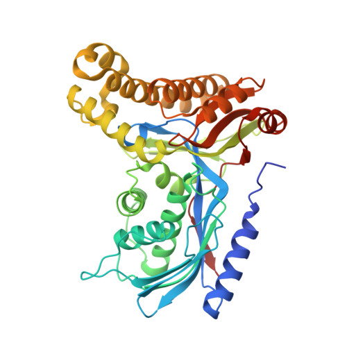
    Classic galactosemia is caused by loss-of-function mutations in galactose-1-phosphate uridylyltransferase (GALT) that lead to toxic accumulation of its substrate, galactose-1-phosphate. One proposed therapy is to inhibit the biosynthesis of galactose-1-phosphate, catalyzed by galactokinase 1 (GALK1). Existing inhibitors of human GALK1 (hGALK1) are primarily ATP-competitive with limited clinical utility to date. Here, we determined crystal structures of hGALK1 bound with reported ATP-competitive inhibitors of the spiro-benzoxazole series, to reveal their binding mode in the active site. Spurred by the need for additional chemotypes of hGALK1 inhibitors, desirably targeting a nonorthosteric site, we also performed crystallography-based screening by soaking hundreds of hGALK1 crystals, already containing active site ligands, with fragments from a custom library. Two fragments were found to bind close to the ATP binding site, and a further eight were found in a hotspot distal from the active site, highlighting the strength of this method in identifying previously uncharacterized allosteric sites. To generate inhibitors of improved potency and selectivity targeting the newly identified binding hotspot, new compounds were designed by merging overlapping fragments. This yielded two micromolar inhibitors of hGALK1 that were not competitive with respect to either substrate (ATP or galactose) and demonstrated good selectivity over hGALK1 homologues, galactokinase 2 and mevalonate kinase. Our findings are therefore the first to demonstrate inhibition of hGALK1 from an allosteric site, with potential for further development of potent and selective inhibitors to provide novel therapeutics for classic galactosemia.


  • Organizational Affiliation

    Structural Genomics Consortium, Nuffield Department of Medicine, University of Oxford, Oxford, United Kingdom, OX3 7DQ.


Macromolecules
Find similar proteins by:  (by identity cutoff)  |  3D Structure
Entity ID: 1
MoleculeChains Sequence LengthOrganismDetailsImage
GalactokinaseA,
B,
C [auth E],
D
399Homo sapiensMutation(s): 2 
Gene Names: GALK1GALK
EC: 2.7.1.6
UniProt & NIH Common Fund Data Resources
Find proteins for P51570 (Homo sapiens)
Explore P51570 
Go to UniProtKB:  P51570
PHAROS:  P51570
GTEx:  ENSG00000108479 
Entity Groups  
Sequence Clusters30% Identity50% Identity70% Identity90% Identity95% Identity100% Identity
UniProt GroupP51570
Sequence Annotations
Expand
  • Reference Sequence
Small Molecules
Ligands 3 Unique
IDChains Name / Formula / InChI Key2D Diagram3D Interactions
HFK
Query on HFK

Download Ideal Coordinates CCD File 
F [auth A],
I [auth B],
L [auth E]
2-(1,3-benzoxazol-2-ylamino)spiro[1,6,7,8-tetrahydroquinazoline-4,1'-cyclohexane]-5-one
C20 H22 N4 O2
AZBGHUMVHJKFOO-UHFFFAOYSA-N
QL5 (Subject of Investigation/LOI)
Query on QL5

Download Ideal Coordinates CCD File 
G [auth A],
J [auth B]
(2,5-dimethylphenyl) pyridine-4-carboxylate
C14 H13 N O2
MMVMRVVLUMIUAF-UHFFFAOYSA-N
GAL
Query on GAL

Download Ideal Coordinates CCD File 
E [auth A],
H [auth B],
K [auth E]
beta-D-galactopyranose
C6 H12 O6
WQZGKKKJIJFFOK-FPRJBGLDSA-N
Experimental Data & Validation

Experimental Data

  • Method: X-RAY DIFFRACTION
  • Resolution: 2.30 Å
  • R-Value Free: 0.254 
  • R-Value Work: 0.206 
  • R-Value Observed: 0.208 
  • Space Group: P 1 21 1
Unit Cell:
Length ( Å )Angle ( ˚ )
a = 73.308α = 90
b = 114.628β = 100.6
c = 120.824γ = 90
Software Package:
Software NamePurpose
XDSdata reduction
Aimlessdata scaling
REFMACrefinement
PDB_EXTRACTdata extraction
REFMACphasing

Structure Validation

View Full Validation Report


Ligand Structure Quality Assessment 


Entry History & Funding Information

Deposition Data


Funding OrganizationLocationGrant Number
Wellcome TrustUnited Kingdom092809/Z/10/Z

Revision History  (Full details and data files)

  • Version 1.0: 2020-07-29
    Type: Initial release
  • Version 1.1: 2021-04-07
    Changes: Database references
  • Version 1.2: 2021-04-28
    Changes: Database references
  • Version 1.3: 2024-01-24
    Changes: Data collection, Database references, Refinement description