6YX1 | pdb_00006yx1

Crystal structure of SHANK1 PDZ in complex with a peptide-small molecule hybrid


Experimental Data Snapshot

  • Method: X-RAY DIFFRACTION
  • Resolution: 1.80 Å
  • R-Value Free: 
    0.255 (Depositor) 
  • R-Value Work: 
    0.222 (Depositor) 
  • R-Value Observed: 
    0.225 (Depositor) 

Starting Model: experimental
View more details

wwPDB Validation 3D Report Full Report

Validation slider image for 6YX1

Ligand Structure Quality Assessment 


This is version 1.2 of the entry. See complete history

Literature

Identification of beta-strand mediated protein-protein interaction inhibitors using ligand-directed fragment ligation.

Hegedus, Z.Hobor, F.Shoemark, D.K.Celis, S.Lian, L.Y.Trinh, C.H.Sessions, R.B.Edwards, T.A.Wilson, A.J.

(2021) Chem Sci 12: 2286-2293

  • DOI: https://doi.org/10.1039/d0sc05694d
  • Primary Citation Related Structures: 
    6YWZ, 6YX0, 6YX1, 6YX2

  • PubMed Abstract: 

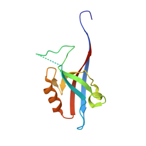
    β-Strand mediated protein-protein interactions (PPIs) represent underexploited targets for chemical probe development despite representing a significant proportion of known and therapeutically relevant PPI targets. β-Strand mimicry is challenging given that both amino acid side-chains and backbone hydrogen-bonds are typically required for molecular recognition, yet these are oriented along perpendicular vectors. This paper describes an alternative approach, using GKAP/SHANK1 PDZ as a model and dynamic ligation screening to identify small-molecule replacements for tranches of peptide sequence. A peptide truncation of GKAP functionalized at the N- and C-termini with acylhydrazone groups was used as an anchor. Reversible acylhydrazone bond exchange with a library of aldehyde fragments in the presence of the protein as template and in situ screening using a fluorescence anisotropy (FA) assay identified peptide hybrid hits with comparable affinity to the GKAP peptide binding sequence. Identified hits were validated using FA, ITC, NMR and X-ray crystallography to confirm selective inhibition of the target PDZ-mediated PPI and mode of binding. These analyses together with molecular dynamics simulations demonstrated the ligands make transient interactions with an unoccupied basic patch through electrostatic interactions, establishing proof-of-concept that this unbiased approach to ligand discovery represents a powerful addition to the armory of tools that can be used to identify PPI modulators.


  • Organizational Affiliation
    • School of Chemistry, University of Leeds Woodhouse Lane Leeds LS2 9JT UK a.j.wilson@leeds.ac.uk.

Macromolecule Content 

  • Total Structure Weight: 26.06 kDa 
  • Atom Count: 1,819 
  • Modeled Residue Count: 210 
  • Deposited Residue Count: 224 
  • Unique protein chains: 1

Macromolecules

Find similar proteins by:|  3D Structure
Entity ID: 1
MoleculeChains  Sequence LengthOrganismDetailsImage
SH3 and multiple ankyrin repeat domains protein 1
A, B
112Homo sapiensMutation(s): 0 
Gene Names: SHANK1
UniProt & NIH Common Fund Data Resources
Find proteins for Q9Y566 (Homo sapiens)
Explore Q9Y566 
Go to UniProtKB:  Q9Y566
PHAROS:  Q9Y566
GTEx:  ENSG00000161681 
Entity Groups
Sequence Clusters30% Identity50% Identity70% Identity90% Identity95% Identity100% Identity
UniProt GroupQ9Y566
Sequence Annotations
Expand
Reference Sequence

Small Molecules

Ligands 4 Unique
IDChains Name / Formula / InChI Key2D Diagram3D Interactions
PWT

Query on PWT


Download:Ideal Coordinates CCD File
C [auth A],
G [auth B]
2-[[2-(5-oxidanylidenepentanoyl)hydrazinyl]methyl]benzoic acid
C13 H16 N2 O4
WAEOIFKXEDZIQC-UHFFFAOYSA-N
ARG

Query on ARG


Download:Ideal Coordinates CCD File
E [auth A],
I [auth B]
ARGININE
C6 H15 N4 O2
ODKSFYDXXFIFQN-BYPYZUCNSA-O
LEU

Query on LEU


Download:Ideal Coordinates CCD File
F [auth A],
J [auth B]
LEUCINE
C6 H13 N O2
ROHFNLRQFUQHCH-YFKPBYRVSA-N
THR

Query on THR


Download:Ideal Coordinates CCD File
D [auth A],
H [auth B]
THREONINE
C4 H9 N O3
AYFVYJQAPQTCCC-GBXIJSLDSA-N

Experimental Data & Validation

Experimental Data

  • Method: X-RAY DIFFRACTION
  • Resolution: 1.80 Å
  • R-Value Free:  0.255 (Depositor) 
  • R-Value Work:  0.222 (Depositor) 
  • R-Value Observed: 0.225 (Depositor) 
Space Group: P 21 21 21
Unit Cell:
Length ( Å )Angle ( ˚ )
a = 44.668α = 90
b = 65.912β = 90
c = 85.637γ = 90
Software Package:
Software NamePurpose
REFMACrefinement
PHENIXrefinement
PDB_EXTRACTdata extraction
STARANISOdata scaling
autoPROCdata scaling
PHASERphasing
Cootmodel building
XDSdata reduction

Structure Validation

View Full Validation Report


Ligand Structure Quality Assessment 


Entry History 

& Funding Information

Deposition Data


Funding OrganizationLocationGrant Number
Engineering and Physical Sciences Research CouncilUnited KingdomEP/N035267/1
European CommissionUnited KingdomMSCA-IF-2016-749012
Royal SocietyUnited KingdomSRF/R1/191087

Revision History  (Full details and data files)

  • Version 1.0: 2021-01-20
    Type: Initial release
  • Version 1.1: 2021-07-07
    Changes: Database references
  • Version 1.2: 2024-01-24
    Changes: Data collection, Database references, Refinement description