6YVJ

EED in complex with a triazolopyrimidine


Experimental Data Snapshot

  • Method: X-RAY DIFFRACTION
  • Resolution: 1.84 Å
  • R-Value Free: 0.199 
  • R-Value Work: 0.182 
  • R-Value Observed: 0.182 

wwPDB Validation   3D Report Full Report


Ligand Structure Quality Assessment 


This is version 2.1 of the entry. See complete history


Literature

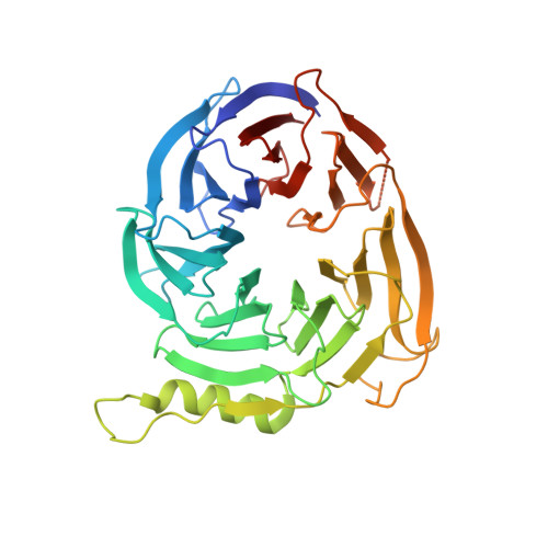
Free energy perturbation in the design of EED ligands as inhibitors of polycomb repressive complex 2 (PRC2) methyltransferase.

O' Donovan, D.H.Gregson, C.Packer, M.J.Greenwood, R.Pike, K.G.Kawatkar, S.Bloecher, A.Robinson, J.Read, J.Code, E.Hsu, J.H.Shen, M.Woods, H.Barton, P.Fillery, S.Williamson, B.Rawlins, P.B.Bagal, S.K.

(2021) Bioorg Med Chem Lett 39: 127904-127904

  • DOI: https://doi.org/10.1016/j.bmcl.2021.127904
  • Primary Citation of Related Structures:  
    6YVI, 6YVJ

  • PubMed Abstract: 

    Free Energy Perturbation (FEP) calculations can provide high-confidence predictions of the interaction strength between a ligand and its protein target. We sought to explore a series of triazolopyrimidines which bind to the EED subunit of the PRC2 complex as potential anticancer therapeutics, using FEP calculations to inform compound design. Combining FEP predictions with a late-stage functionalisation (LSF) inspired synthetic approach allowed us to rapidly evaluate structural modifications in a previously unexplored region of the EED binding site. This approach generated a series of novel triazolopyrimidine EED ligands with improved physicochemical properties and which inhibit PRC2 methyltransferase activity in a cancer-relevant G401 cell line.


  • Organizational Affiliation

    Oncology R&D, AstraZeneca, Cambridge CB4 0WG, United Kingdom. Electronic address: daniel.odonovan@astrazeneca.com.


Macromolecules
Find similar proteins by:  (by identity cutoff)  |  3D Structure
Entity ID: 1
MoleculeChains Sequence LengthOrganismDetailsImage
Polycomb protein EED
A, B
366Homo sapiensMutation(s): 0 
Gene Names: EED
UniProt & NIH Common Fund Data Resources
Find proteins for O75530 (Homo sapiens)
Explore O75530 
Go to UniProtKB:  O75530
PHAROS:  O75530
GTEx:  ENSG00000074266 
Entity Groups  
Sequence Clusters30% Identity50% Identity70% Identity90% Identity95% Identity100% Identity
UniProt GroupO75530
Sequence Annotations
Expand
  • Reference Sequence
Small Molecules
Ligands 3 Unique
IDChains Name / Formula / InChI Key2D Diagram3D Interactions
L9W
Query on L9W

Download Ideal Coordinates CCD File 
C [auth A]N-(2,3-dihydro-1-benzofuran-4-ylmethyl)-8-(4-methylsulfonylphenyl)-[1,2,4]triazolo[4,3-c]pyrimidin-5-amine
C21 H19 N5 O3 S
GDDMEOORUYUIJL-UHFFFAOYSA-N
EJR (Subject of Investigation/LOI)
Query on EJR

Download Ideal Coordinates CCD File 
E [auth B]N-[(5-fluoranyl-2,3-dihydro-1-benzofuran-4-yl)methyl]-8-(2-methylpyridin-3-yl)-[1,2,4]triazolo[4,3-c]pyrimidin-5-amine
C20 H17 F N6 O
XLIBABIFOBYHSV-UHFFFAOYSA-N
GOL
Query on GOL

Download Ideal Coordinates CCD File 
D [auth B]GLYCEROL
C3 H8 O3
PEDCQBHIVMGVHV-UHFFFAOYSA-N
Experimental Data & Validation

Experimental Data

  • Method: X-RAY DIFFRACTION
  • Resolution: 1.84 Å
  • R-Value Free: 0.199 
  • R-Value Work: 0.182 
  • R-Value Observed: 0.182 
  • Space Group: P 21 21 21
Unit Cell:
Length ( Å )Angle ( ˚ )
a = 87.02α = 90
b = 89.87β = 90
c = 99.14γ = 90
Software Package:
Software NamePurpose
Aimlessdata scaling
BUSTERrefinement
PDB_EXTRACTdata extraction
XDSdata reduction
AMoREphasing

Structure Validation

View Full Validation Report


Ligand Structure Quality Assessment 


Entry History 

Deposition Data

  • Released Date: 2021-05-12 
  • Deposition Author(s): Read, J.A.

Revision History  (Full details and data files)

  • Version 1.0: 2021-05-12
    Type: Initial release
  • Version 2.0: 2021-07-14
    Changes: Database references, Derived calculations, Non-polymer description, Refinement description, Structure summary
  • Version 2.1: 2024-01-24
    Changes: Data collection, Database references, Refinement description