6YNA

Crystal structure of cAMP-dependent Protein Kinase (PKA) in complex with Fasudil (M77, soaked)


Experimental Data Snapshot

  • Method: X-RAY DIFFRACTION
  • Resolution: 1.47 Å
  • R-Value Free: 0.189 
  • R-Value Work: 0.163 
  • R-Value Observed: 0.165 

wwPDB Validation   3D Report Full Report


Ligand Structure Quality Assessment 


This is version 1.3 of the entry. See complete history


Literature

Two Methods, One Goal: Structural Differences between Cocrystallization and Crystal Soaking to Discover Ligand Binding Poses.

Wienen-Schmidt, B.Oebbeke, M.Ngo, K.Heine, A.Klebe, G.

(2021) ChemMedChem 16: 292-300

  • DOI: https://doi.org/10.1002/cmdc.202000565
  • Primary Citation of Related Structures:  
    6YNA, 6YNB, 6YNC, 6YNR, 6YNT, 6YQI, 6YQJ, 6YQK

  • PubMed Abstract: 

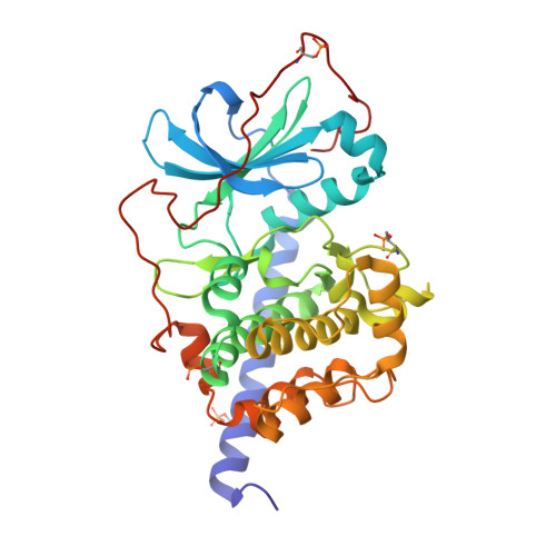
    In lead optimization, protein crystallography is an indispensable tool to analyze drug binding. Binding modes and non-covalent interaction inventories are essential to design follow-up synthesis candidates. Two protocols are commonly applied to produce protein-ligand complexes: cocrystallization and soaking. Because of its time and cost effectiveness, soaking is the more popular method. Taking eight ligand hinge binders of protein kinase A, we demonstrate that cocrystallization is superior. Particularly for flexible proteins, such as kinases, and larger ligands cocrystallization captures more reliable the correct binding pose and induced protein adaptations. The geometrical discrepancies between soaking and cocrystallization appear smaller for fragment-sized ligands. For larger flexible ligands that trigger conformational changes of the protein, soaking can be misleading and underestimates the number of possible polar interactions due to inadequate, highly impaired positions of protein amino-acid side and main chain atoms. Thus, if applicable cocrystallization should be the gold standard to study protein-ligand complexes.


  • Organizational Affiliation

    Institut für Pharmazeutische Chemie, Philipps-Universität Marburg, Marbacher Weg 6, 35032, Marburg, Germany.


Macromolecules
Find similar proteins by:  (by identity cutoff)  |  3D Structure
Entity ID: 1
MoleculeChains Sequence LengthOrganismDetailsImage
cAMP-dependent protein kinase catalytic subunit alpha353Cricetulus griseusMutation(s): 0 
Gene Names: PRKACA
EC: 2.7.11.11
UniProt
Find proteins for P25321 (Cricetulus griseus)
Explore P25321 
Go to UniProtKB:  P25321
Entity Groups  
Sequence Clusters30% Identity50% Identity70% Identity90% Identity95% Identity100% Identity
UniProt GroupP25321
Sequence Annotations
Expand
  • Reference Sequence

Find similar proteins by:  Sequence   |   3D Structure  

Entity ID: 2
MoleculeChains Sequence LengthOrganismDetailsImage
cAMP-dependent protein kinase inhibitor alpha19Mus musculusMutation(s): 4 
UniProt & NIH Common Fund Data Resources
Find proteins for P63248 (Mus musculus)
Explore P63248 
Go to UniProtKB:  P63248
IMPC:  MGI:104747
Entity Groups  
Sequence Clusters30% Identity50% Identity70% Identity90% Identity95% Identity100% Identity
UniProt GroupP63248
Sequence Annotations
Expand
  • Reference Sequence
Small Molecules
Ligands 2 Unique
IDChains Name / Formula / InChI Key2D Diagram3D Interactions
M77 (Subject of Investigation/LOI)
Query on M77

Download Ideal Coordinates CCD File 
D [auth A]5-(1,4-DIAZEPAN-1-SULFONYL)ISOQUINOLINE
C14 H17 N3 O2 S
NGOGFTYYXHNFQH-UHFFFAOYSA-N
MPD
Query on MPD

Download Ideal Coordinates CCD File 
C [auth A](4S)-2-METHYL-2,4-PENTANEDIOL
C6 H14 O2
SVTBMSDMJJWYQN-YFKPBYRVSA-N
Modified Residues  2 Unique
IDChains TypeFormula2D DiagramParent
SEP
Query on SEP
A
L-PEPTIDE LINKINGC3 H8 N O6 PSER
TPO
Query on TPO
A
L-PEPTIDE LINKINGC4 H10 N O6 PTHR
Experimental Data & Validation

Experimental Data

  • Method: X-RAY DIFFRACTION
  • Resolution: 1.47 Å
  • R-Value Free: 0.189 
  • R-Value Work: 0.163 
  • R-Value Observed: 0.165 
  • Space Group: P 21 21 21
Unit Cell:
Length ( Å )Angle ( ˚ )
a = 58.313α = 90
b = 72.547β = 90
c = 109.039γ = 90
Software Package:
Software NamePurpose
PHASERphasing
PHENIXrefinement
XDSdata reduction
XDSdata scaling
Cootmodel building

Structure Validation

View Full Validation Report


Ligand Structure Quality Assessment 


Entry History 

Deposition Data

Revision History  (Full details and data files)

  • Version 1.0: 2020-10-14
    Type: Initial release
  • Version 1.1: 2020-10-21
    Changes: Database references
  • Version 1.2: 2021-01-20
    Changes: Database references
  • Version 1.3: 2024-01-24
    Changes: Data collection, Database references, Refinement description