6YEI

Arabidopsis thaliana glutamate dehydrogenase isoform 1 in complex with NAD


Experimental Data Snapshot

  • Method: X-RAY DIFFRACTION
  • Resolution: 2.02 Å
  • R-Value Free: 0.196 
  • R-Value Work: 0.160 
  • R-Value Observed: 0.161 

wwPDB Validation   3D Report Full Report


Ligand Structure Quality Assessment 


This is version 1.2 of the entry. See complete history


Literature

Structural Studies of Glutamate Dehydrogenase (Isoform 1) FromArabidopsis thaliana, an Important Enzyme at the Branch-Point Between Carbon and Nitrogen Metabolism.

Grzechowiak, M.Sliwiak, J.Jaskolski, M.Ruszkowski, M.

(2020) Front Plant Sci 11: 754-754

  • DOI: https://doi.org/10.3389/fpls.2020.00754
  • Primary Citation of Related Structures:  
    6YEH, 6YEI

  • PubMed Abstract: 

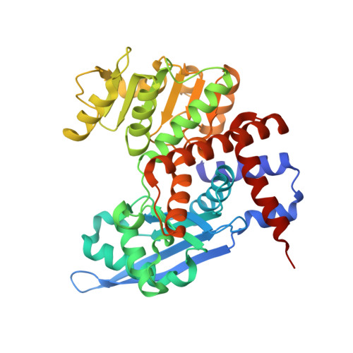
    Glutamate dehydrogenase (GDH) releases ammonia in a reversible NAD(P) + -dependent oxidative deamination of glutamate that yields 2-oxoglutarate (2OG). In current perception, GDH contributes to Glu homeostasis and plays a significant role at the junction of carbon and nitrogen assimilation pathways. GDHs are members of a superfamily of ELFV (Glu/Leu/Phe/Val) amino acid dehydrogenases and are subdivided into three subclasses, based on coenzyme specificity: NAD + -specific, NAD + /NADP + dual-specific, and NADP + -specific. We determined in this work that the mitochondrial At GDH1 isozyme from A. thaliana is NAD + -specific. Altogether, A. thaliana expresses three GDH isozymes ( At GDH1-3) targeted to mitochondria, of which At GDH2 has an extra EF-hand motif and is stimulated by calcium. Our enzymatic assays of At GDH1 established that its sensitivity to calcium is negligible. In vivo the At GDH1-3 enzymes form homo- and heterohexamers of varied composition. We solved the crystal structure of recombinant At GDH1 in the apo-form and in complex with NAD + at 2.59 and 2.03 Å resolution, respectively. We demonstrate also that both in the apo form and in 1:1 complex with NAD + , it forms D 3 -symmetric homohexamers. A subunit of At GDH1 consists of domain I, which is involved in hexamer formation and substrate binding, and of domain II which binds coenzyme. Most of the subunits in our crystal structures, including those in NAD + complex, are in open conformation, with domain II forming a large (albeit variable) angle with domain I. One of the subunits of the At GDH1-NAD + hexamer contains a serendipitous 2OG molecule in the active site, causing a dramatic (∼25°) closure of the domains. We provide convincing evidence that the N-terminal peptide preceding domain I is a mitochondrial targeting signal, with a predicted cleavage site for mitochondrial processing peptidase (MPP) at Leu17-Leu18 that is followed by an unexpected potassium coordination site (Ser27, Ile30). We also identified several MPD [(+/-)-2-methyl-2,4-pentanediol] binding sites with conserved sequence. Although At GDH1 is insensitive to MPD in our assays, the observation of druggable sites opens a potential for non-competitive herbicide design.


  • Organizational Affiliation

    Center for Biocrystallographic Research Institute of Bioorganic Chemistry, Polish Academy of Sciences, Poznań, Poland.


Macromolecules
Find similar proteins by:  (by identity cutoff)  |  3D Structure
Entity ID: 1
MoleculeChains Sequence LengthOrganismDetailsImage
Glutamate dehydrogenase 1
A, B, C, D, E
A, B, C, D, E, F
414Arabidopsis thalianaMutation(s): 0 
Gene Names: GDH1At5g18170MRG7.13
EC: 1.4.1.3
UniProt
Find proteins for Q43314 (Arabidopsis thaliana)
Explore Q43314 
Go to UniProtKB:  Q43314
Entity Groups  
Sequence Clusters30% Identity50% Identity70% Identity90% Identity95% Identity100% Identity
UniProt GroupQ43314
Sequence Annotations
Expand
  • Reference Sequence
Small Molecules
Ligands 6 Unique
IDChains Name / Formula / InChI Key2D Diagram3D Interactions
NAD (Subject of Investigation/LOI)
Query on NAD

Download Ideal Coordinates CCD File 
G [auth A]
HA [auth E]
MA [auth F]
O [auth B]
T [auth C]
G [auth A],
HA [auth E],
MA [auth F],
O [auth B],
T [auth C],
Z [auth D]
NICOTINAMIDE-ADENINE-DINUCLEOTIDE
C21 H27 N7 O14 P2
BAWFJGJZGIEFAR-NNYOXOHSSA-N
AKG (Subject of Investigation/LOI)
Query on AKG

Download Ideal Coordinates CCD File 
NA [auth F]2-OXOGLUTARIC ACID
C5 H6 O5
KPGXRSRHYNQIFN-UHFFFAOYSA-N
MRD
Query on MRD

Download Ideal Coordinates CCD File 
FA [auth D],
KA [auth E],
N [auth A],
S [auth B],
W [auth C]
(4R)-2-METHYLPENTANE-2,4-DIOL
C6 H14 O2
SVTBMSDMJJWYQN-RXMQYKEDSA-N
MPD
Query on MPD

Download Ideal Coordinates CCD File 
AA [auth D]
BA [auth D]
CA [auth D]
DA [auth D]
GA [auth E]
AA [auth D],
BA [auth D],
CA [auth D],
DA [auth D],
GA [auth E],
H [auth A],
I [auth A],
IA [auth E],
J [auth A],
K [auth A],
P [auth B],
Q [auth B],
R [auth B],
U [auth C],
V [auth C]
(4S)-2-METHYL-2,4-PENTANEDIOL
C6 H14 O2
SVTBMSDMJJWYQN-YFKPBYRVSA-N
EDO
Query on EDO

Download Ideal Coordinates CCD File 
OA [auth F],
Y [auth D]
1,2-ETHANEDIOL
C2 H6 O2
LYCAIKOWRPUZTN-UHFFFAOYSA-N
K (Subject of Investigation/LOI)
Query on K

Download Ideal Coordinates CCD File 
EA [auth D]
JA [auth E]
L [auth A]
LA [auth E]
M [auth A]
EA [auth D],
JA [auth E],
L [auth A],
LA [auth E],
M [auth A],
X [auth C]
POTASSIUM ION
K
NPYPAHLBTDXSSS-UHFFFAOYSA-N
Binding Affinity Annotations 
IDSourceBinding Affinity
NAD Binding MOAD:  6YEI Kd: 7.20e+4 (nM) from 1 assay(s)
Experimental Data & Validation

Experimental Data

Unit Cell:
Length ( Å )Angle ( ˚ )
a = 93.843α = 90
b = 99.811β = 90
c = 317.948γ = 90
Software Package:
Software NamePurpose
PHENIXrefinement
XDSdata reduction
XSCALEdata scaling
PHASERphasing

Structure Validation

View Full Validation Report


Ligand Structure Quality Assessment 


Entry History & Funding Information

Deposition Data


Funding OrganizationLocationGrant Number
Polish National Science CentrePolandSONATA 2018/31/D/NZ1/03630

Revision History  (Full details and data files)

  • Version 1.0: 2020-05-20
    Type: Initial release
  • Version 1.1: 2020-07-22
    Changes: Database references
  • Version 1.2: 2024-01-24
    Changes: Data collection, Database references, Derived calculations, Refinement description