
 Download Mendeley
Fucosylated antimicrobial linear peptide cFucRH46D in complex with the fucose binding lectin LecB from Pseudomonas aeruginosa at 2.1 Angstrom resolution
Baeriswyl, S., Stocker, A., Reymond, J.-L.To be published.
Experimental Data Snapshot
Starting Model: experimental
View more details
Entity ID: 1 | |||||
|---|---|---|---|---|---|
| Molecule | Chains | Sequence Length | Organism | Details | Image |
| Fucose-binding lectin | A, C [auth B] | 115 | Pseudomonas aeruginosa | Mutation(s): 0 Gene Names: lecB, C0044_25260, CAZ10_21840, DT376_00595, DY979_15445, ECC04_10105, EFK27_13700, EGV95_09240, EGY23_15550, IPC669_23070... | 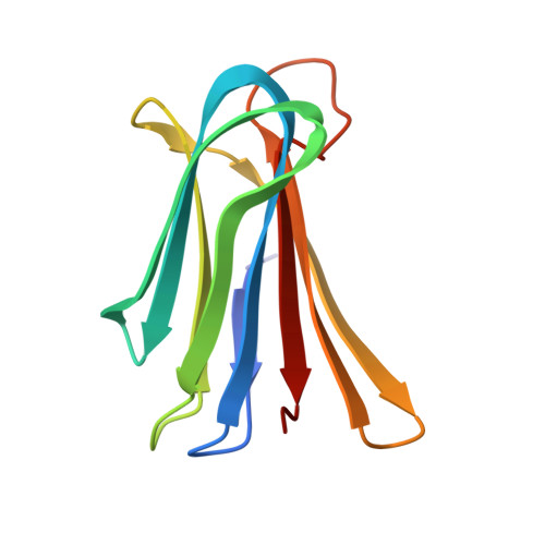 |
UniProt | |||||
Entity Groups | |||||
| Sequence Clusters | 30% Identity50% Identity70% Identity90% Identity95% Identity100% Identity | ||||
| UniProt Group | Q9HYN5 | ||||
Sequence AnnotationsExpand | |||||
Reference Sequence | |||||
Entity ID: 2 | |||||
|---|---|---|---|---|---|
| Molecule | Chains | Sequence Length | Organism | Details | Image |
| cFucRH46D | B [auth C], D | 14 | synthetic construct | Mutation(s): 0 | 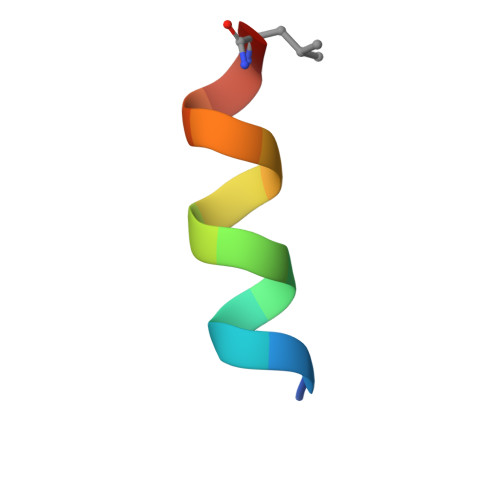 |
Sequence AnnotationsExpand | |||||
Reference Sequence | |||||
| Ligands 2 Unique | |||||
|---|---|---|---|---|---|
| ID | Chains | Name / Formula / InChI Key | 2D Diagram | 3D Interactions | |
| ZDC (Subject of Investigation/LOI) Download:Ideal Coordinates CCD File | G [auth C], J [auth D] | 3,7-anhydro-2,8-dideoxy-L-glycero-D-gluco-octonic acid C8 H14 O6 YTZUDUWVQZSKNN-OASCRQMUSA-N | 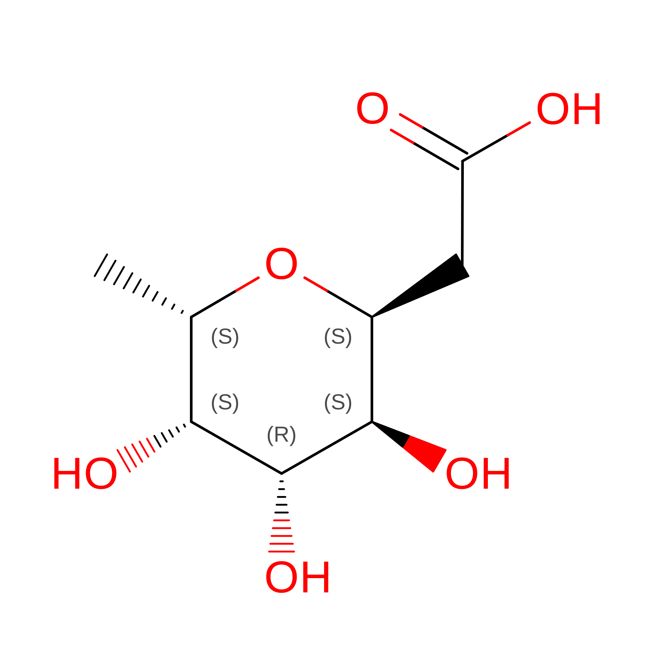 | ||
| CA Download:Ideal Coordinates CCD File | E [auth A], F [auth A], H [auth B], I [auth B] | CALCIUM ION Ca BHPQYMZQTOCNFJ-UHFFFAOYSA-N |  | ||
| Non-standard Monomers 7 Unique | ||||
|---|---|---|---|---|
| ID | Chains | Type | Formula | 2D Diagram |
| DAL Query on DAL | D-PEPTIDE LINKING | C3 H7 N O2 | 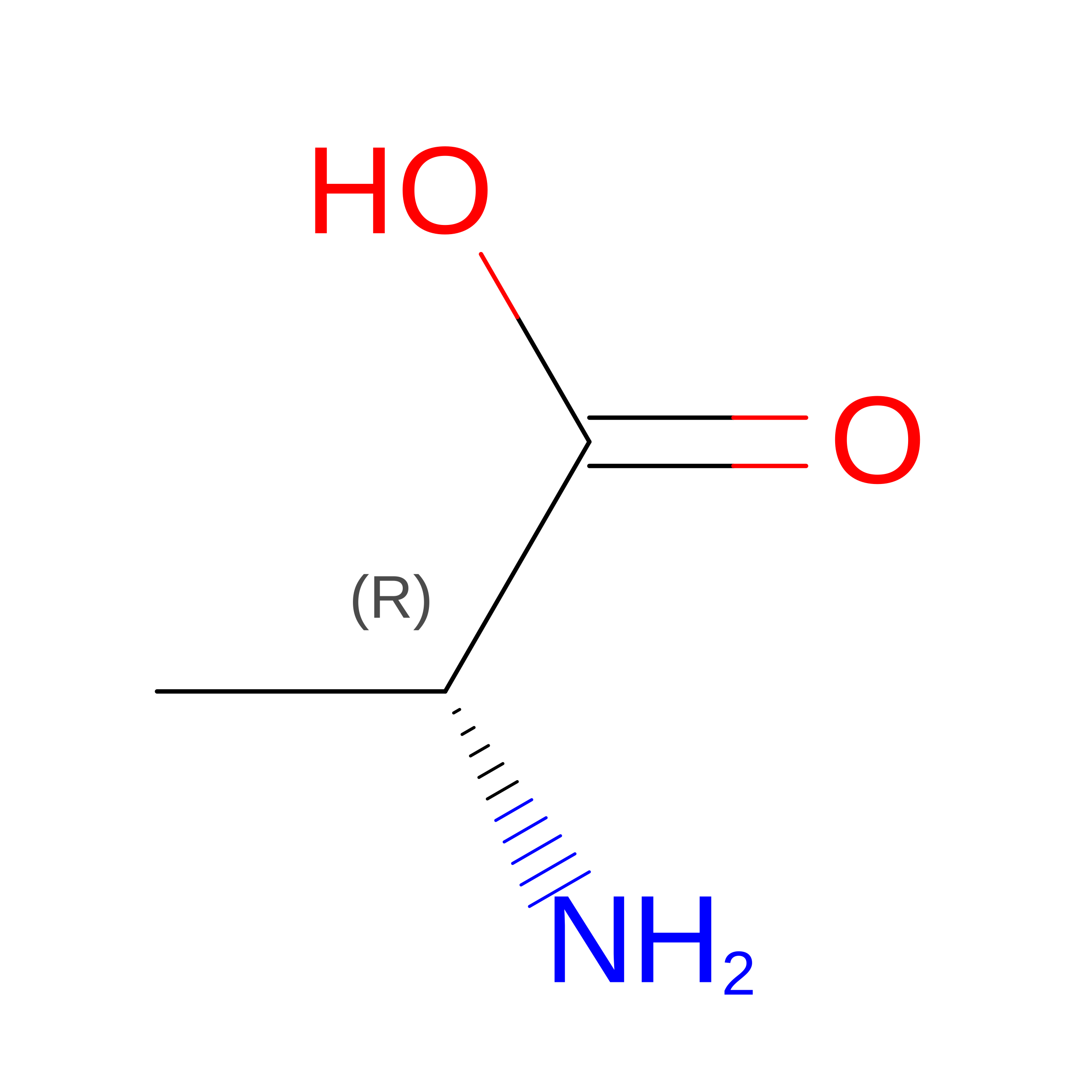 | |
| DHI Query on DHI | D-PEPTIDE LINKING | C6 H10 N3 O2 | 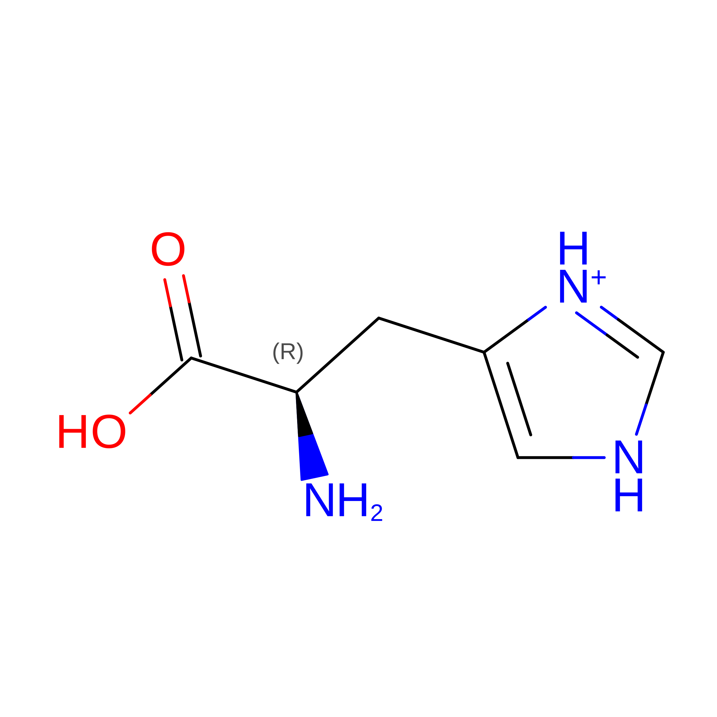 | |
| DLE Query on DLE | D-PEPTIDE LINKING | C6 H13 N O2 | 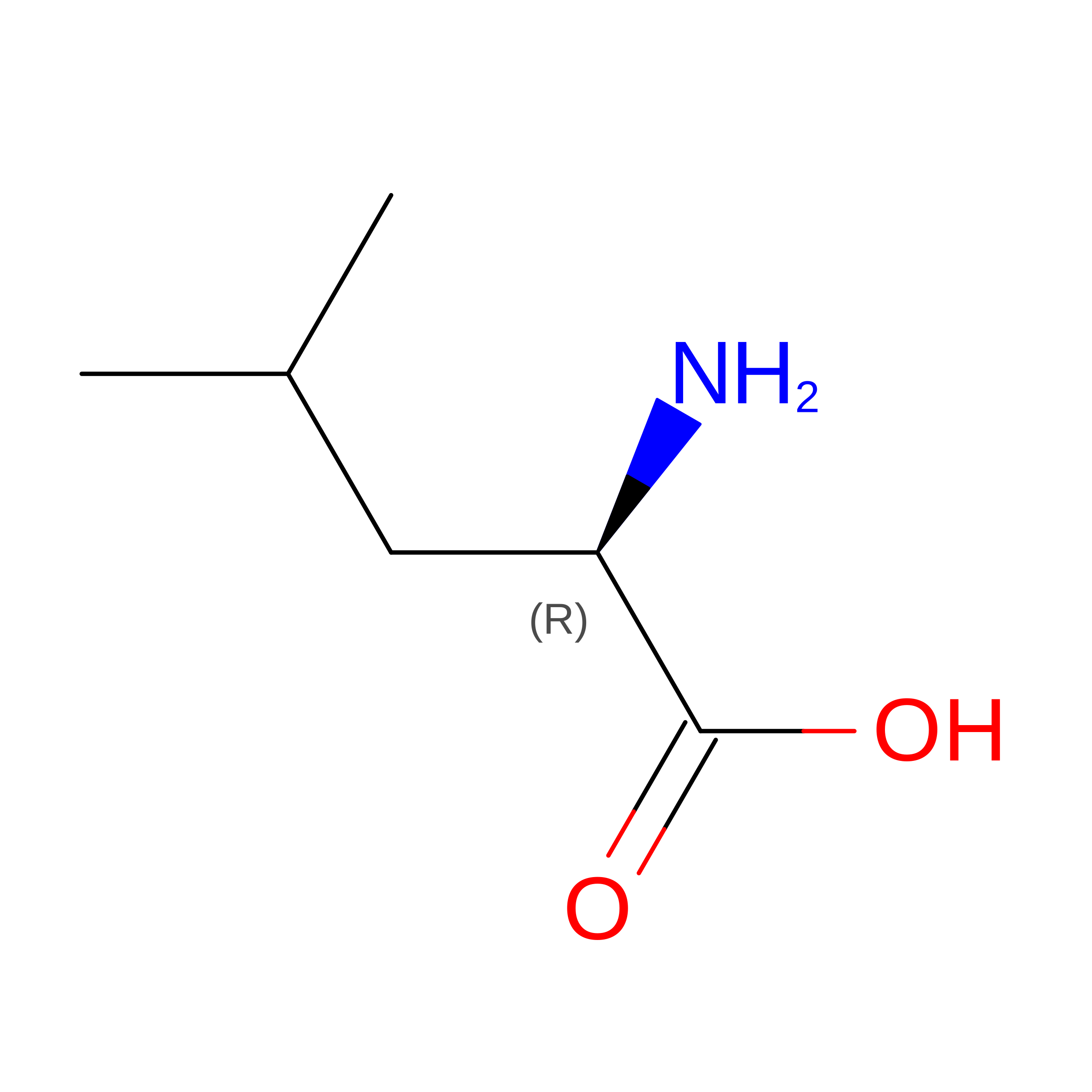 | |
| DLY Query on DLY | D-PEPTIDE LINKING | C6 H14 N2 O2 | 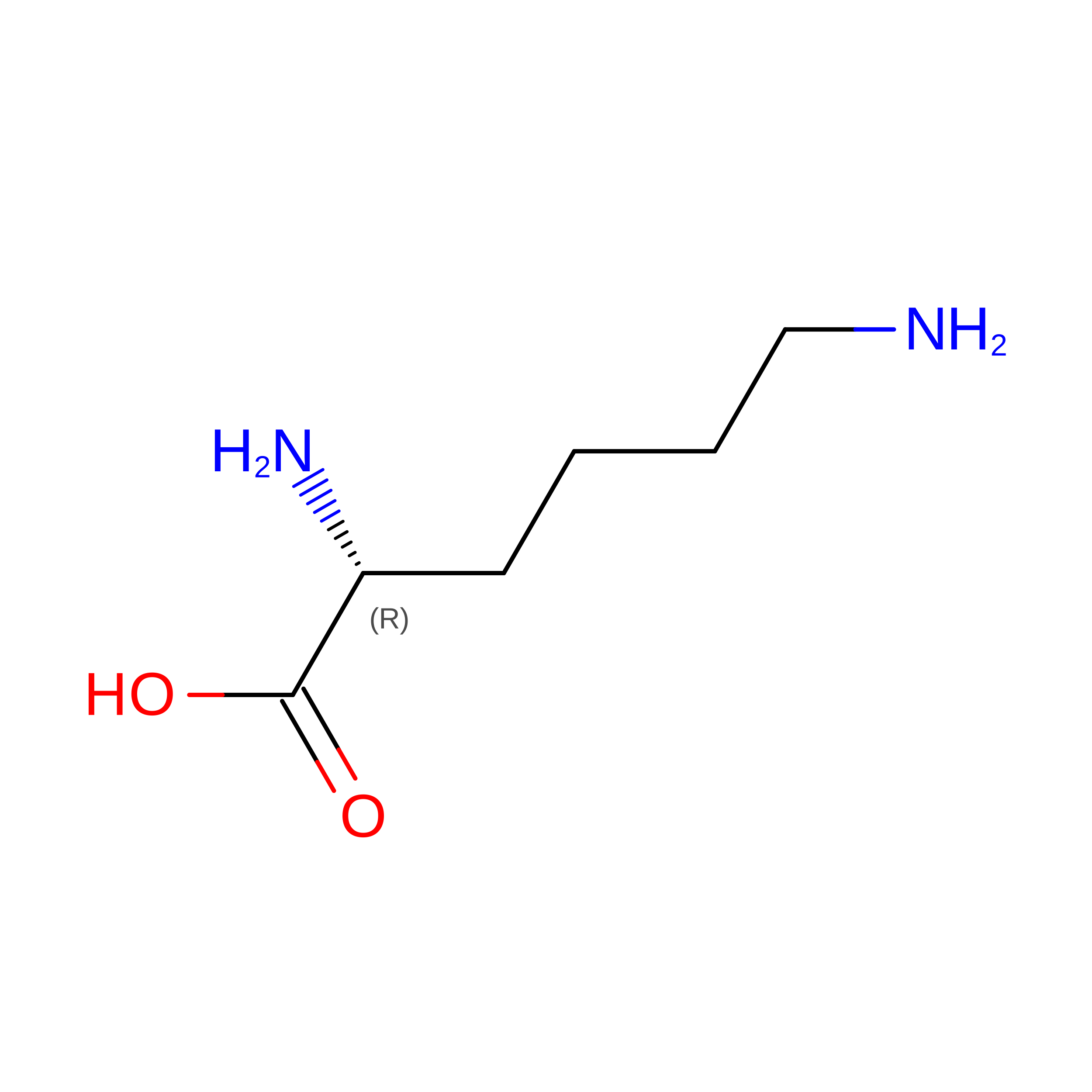 | |
| DPN Query on DPN | D-PEPTIDE LINKING | C9 H11 N O2 | 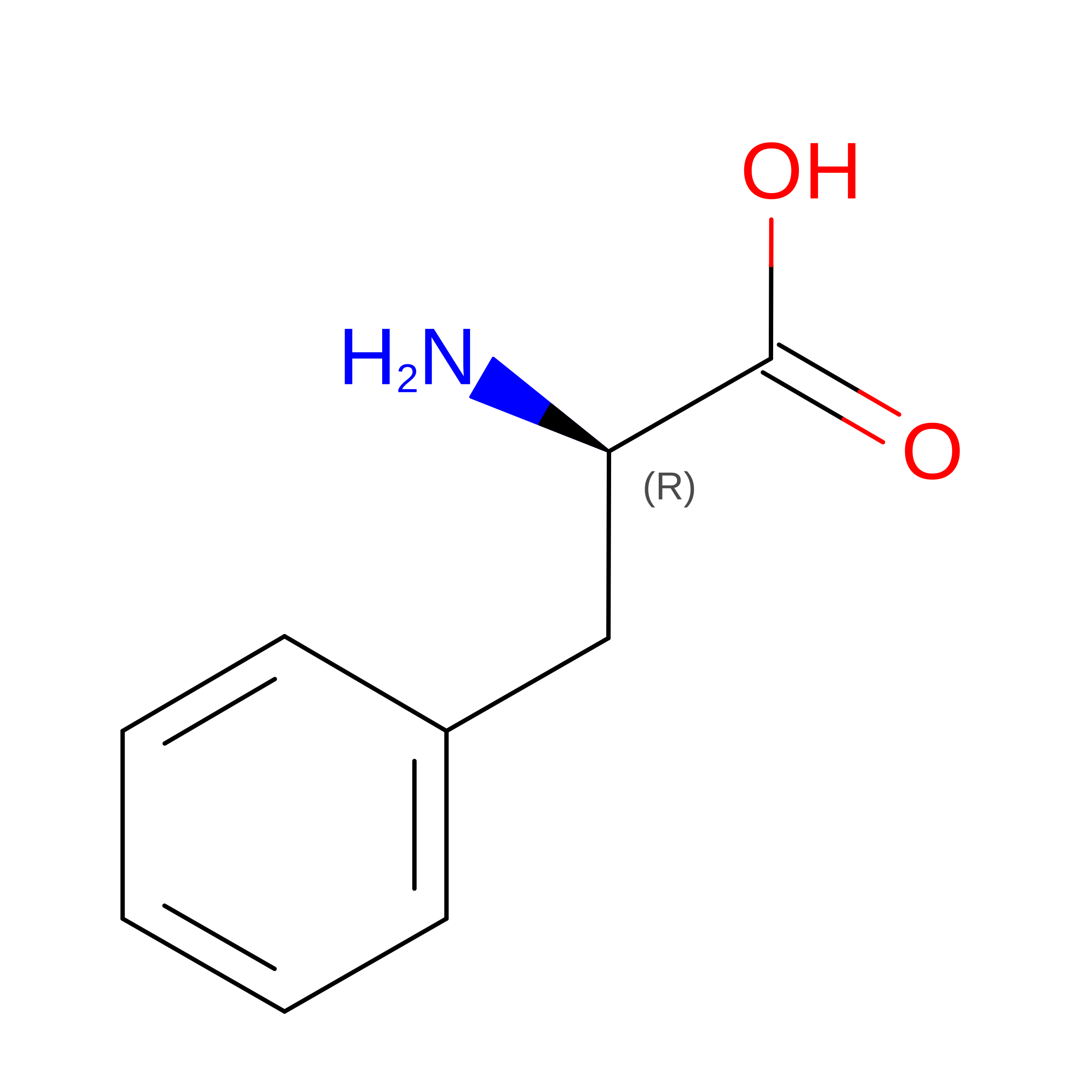 | |
| DTR Query on DTR | D-PEPTIDE LINKING | C11 H12 N2 O2 | 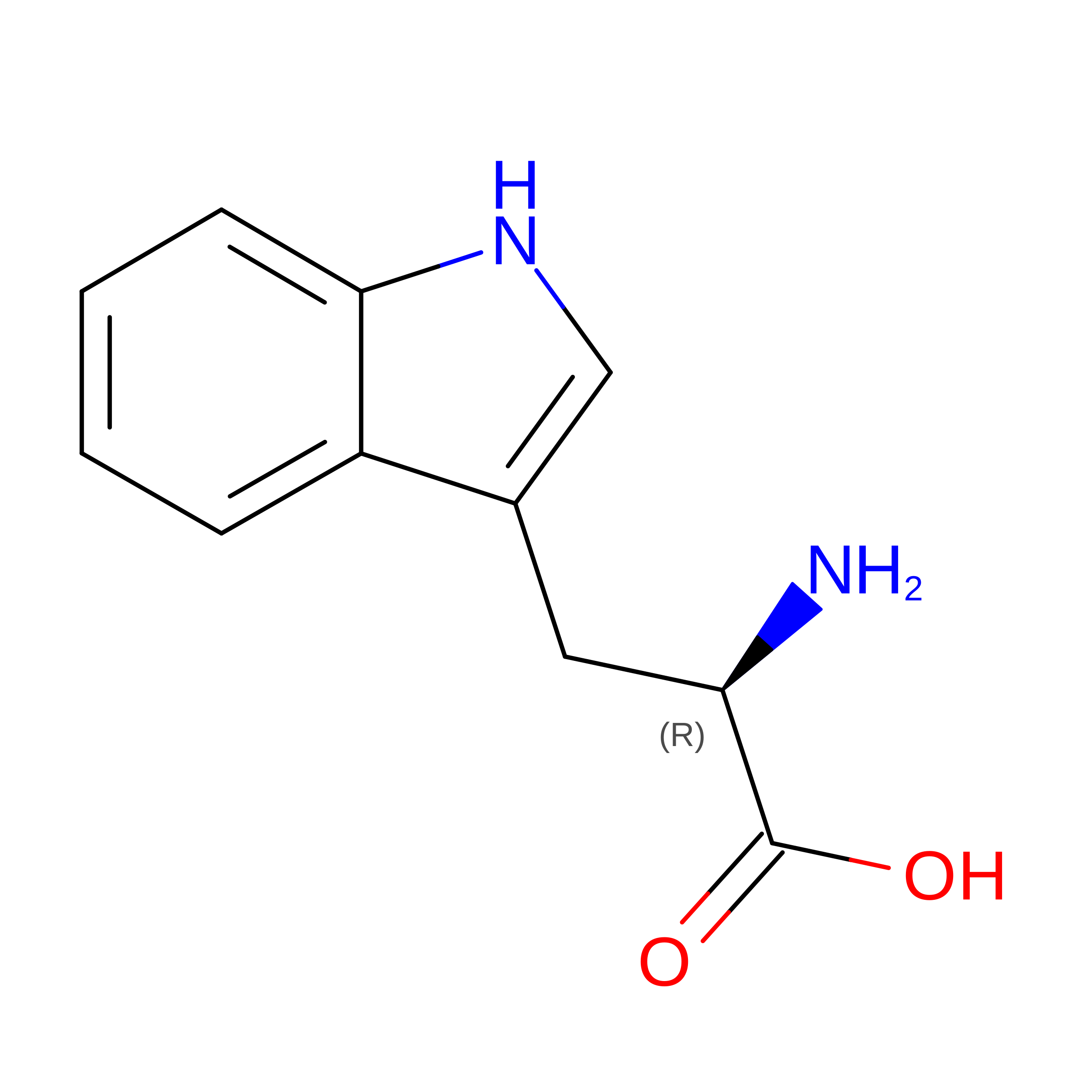 | |
| DTY Query on DTY | D-PEPTIDE LINKING | C9 H11 N O3 | 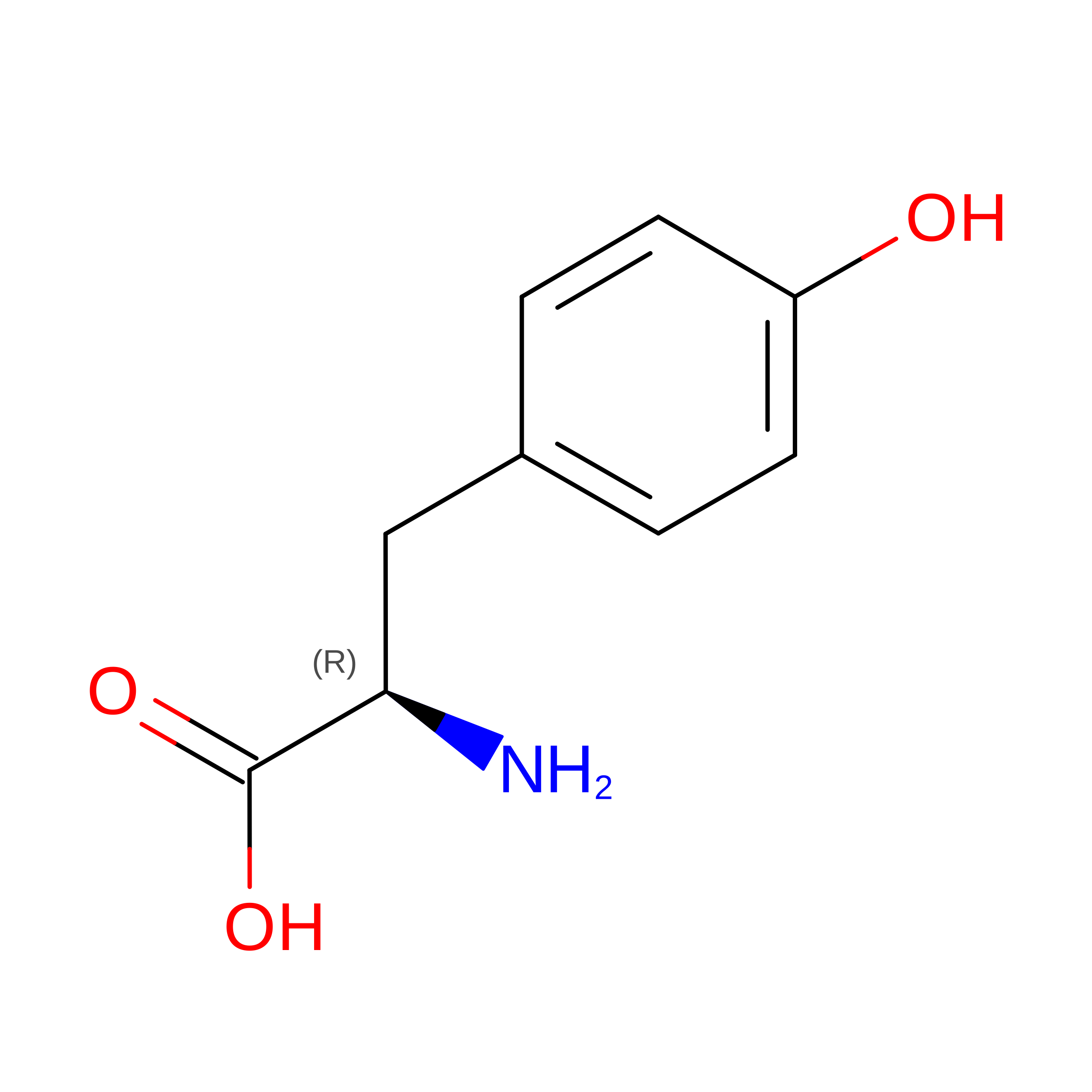 | |
| Length ( Å ) | Angle ( ˚ ) |
|---|---|
| a = 68.999 | α = 90 |
| b = 68.999 | β = 90 |
| c = 271.453 | γ = 120 |
| Software Name | Purpose |
|---|---|
| PHENIX | refinement |
| XDS | data reduction |
| SCALA | data scaling |
| PHASER | phasing |
| PDB_EXTRACT | data extraction |
| Funding Organization | Location | Grant Number |
|---|---|---|
| Swiss National Science Foundation | Switzerland | -- |
