A Single Second Shell Amino Acid Determines Affinity and Kinetics of Linagliptin Binding to Type 4 Dipeptidyl Peptidase and Fibroblast Activation Protein.
Schnapp, G., Hoevels, Y., Bakker, R.A., Schreiner, P., Klein, T., Nar, H.(2021) ChemMedChem 16: 630-639
- PubMed: 33030297 Search on PubMedSearch on PubMed Central
- DOI: https://doi.org/10.1002/cmdc.202000591
- Primary Citation Related Structures:
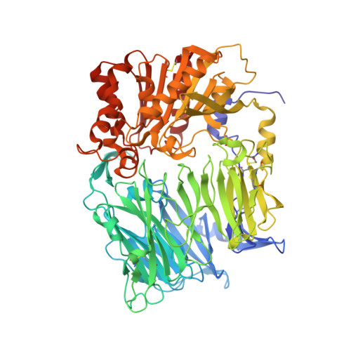
6Y0F - PubMed Abstract:
Drugs targeting type 4 dipeptidyl peptidase (DPP-4) are beneficial for glycemic control, whereas fibroblast activation protein alpha (FAP-α) is a potential target for cancer therapies. Unlike other gliptins, linagliptin displays FAP inhibition. We compared biophysical and structural characteristics of linagliptin binding to DPP-4 and FAP to better understand what differentiates linagliptin from other gliptins. Linagliptin exhibited high binding affinity (K D ) and a slow off-rate (k off ) when dissociating from DPP-4 (K D 6.6 pM; k off 5.1×10 -5 s -1 ), and weaker inhibitory potency to FAP (K D 301 nM; k off >1 s -1 ). Co-structures of linagliptin with DPP-4 or FAP were similar except for one second shell amino acid difference: Asp663 (DPP-4) and Ala657 (FAP). pH dependence of enzymatic activities and binding of linagliptin for DPP-4 and FAP are dependent on this single amino acid difference. While linagliptin may not display any anticancer activity at therapeutic doses, our findings may guide future studies for the development of optimized inhibitors.
- Department of Medicinal Chemistry, Boehringer Ingelheim Pharma GmbH & Co. KG, Birkendorferstr. 65, 88397, Biberach, Germany.
Organizational Affiliation: