6XWS | pdb_00006xws

Crystal Structure of Drosophila CAL1 1-160 bound to CENP-A/H4


Experimental Data Snapshot

  • Method: X-RAY DIFFRACTION
  • Resolution: 4.36 Å
  • R-Value Free: 
    0.323 (Depositor), 0.331 (DCC) 
  • R-Value Work: 
    0.306 (Depositor), 0.305 (DCC) 
  • R-Value Observed: 
    0.307 (Depositor) 

Starting Model: experimental
View more details

wwPDB Validation 3D Report Full Report

Validation slider image for 6XWS

This is version 1.1 of the entry. See complete history

Literature

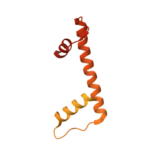
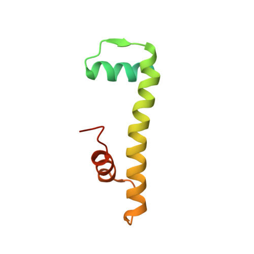
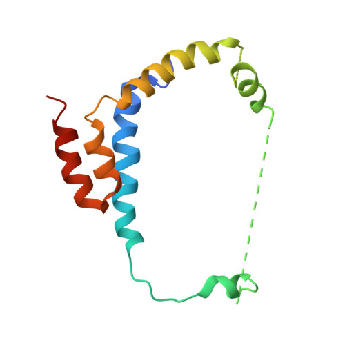
Structural basis for centromere maintenance by Drosophila CENP-A chaperone CAL1.

Medina-Pritchard, B.Lazou, V.Zou, J.Byron, O.Abad, M.A.Rappsilber, J.Heun, P.Jeyaprakash, A.A.

(2020) EMBO J 39: e103234-e103234

  • DOI: https://doi.org/10.15252/embj.2019103234
  • Primary Citation Related Structures: 
    6XWS, 6XWT, 6XWU, 6XWV

  • PubMed Abstract: 

    Centromeres are microtubule attachment sites on chromosomes defined by the enrichment of histone variant CENP-A-containing nucleosomes. To preserve centromere identity, CENP-A must be escorted to centromeres by a CENP-A-specific chaperone for deposition. Despite this essential requirement, many eukaryotes differ in the composition of players involved in centromere maintenance, highlighting the plasticity of this process. In humans, CENP-A recognition and centromere targeting are achieved by HJURP and the Mis18 complex, respectively. Using X-ray crystallography, we here show how Drosophila CAL1, an evolutionarily distinct CENP-A histone chaperone, binds both CENP-A and the centromere receptor CENP-C without the requirement for the Mis18 complex. While an N-terminal CAL1 fragment wraps around CENP-A/H4 through multiple physical contacts, a C-terminal CAL1 fragment directly binds a CENP-C cupin domain dimer. Although divergent at the primary structure level, CAL1 thus binds CENP-A/H4 using evolutionarily conserved and adaptive structural principles. The CAL1 binding site on CENP-C is strategically positioned near the cupin dimerisation interface, restricting binding to just one CAL1 molecule per CENP-C dimer. Overall, by demonstrating how CAL1 binds CENP-A/H4 and CENP-C, we provide key insights into the minimalistic principles underlying centromere maintenance.


  • Organizational Affiliation
    • Wellcome Centre for Cell Biology, University of Edinburgh, Edinburgh, UK.

Macromolecule Content 

  • Total Structure Weight: 47.92 kDa 
  • Atom Count: 1,715 
  • Modeled Residue Count: 242 
  • Deposited Residue Count: 436 
  • Unique protein chains: 3

Macromolecules

Find similar proteins by:|  3D Structure
Entity ID: 1
MoleculeChains  Sequence LengthOrganismDetailsImage
Histone H3-like centromeric protein cid225Drosophila melanogasterMutation(s): 0 
Gene Names: cidCENP-ACG13329
UniProt
Find proteins for Q9V6Q2 (Drosophila melanogaster)
Explore Q9V6Q2 
Go to UniProtKB:  Q9V6Q2
Entity Groups
Sequence Clusters30% Identity50% Identity70% Identity90% Identity95% Identity100% Identity
UniProt GroupQ9V6Q2
Sequence Annotations
Expand
Reference Sequence
Find similar proteins by:|  3D Structure
Entity ID: 2
MoleculeChains  Sequence LengthOrganismDetailsImage
Histone H4103Drosophila melanogasterMutation(s): 0 
Gene Names: His4rBcDNA:RH52884Dmel\CG3379FBtr0082962H4rHis4-88CDHis4RCG3379Dmel_CG3379
UniProt
Find proteins for P84040 (Drosophila melanogaster)
Explore P84040 
Go to UniProtKB:  P84040
Entity Groups
Sequence Clusters30% Identity50% Identity70% Identity90% Identity95% Identity100% Identity
UniProt GroupP84040
Sequence Annotations
Expand
Reference Sequence
Find similar proteins by:|  3D Structure
Entity ID: 3
MoleculeChains  Sequence LengthOrganismDetailsImage
Chromosome alignment defect 1,Chromosome alignment defect 1108Drosophila melanogasterMutation(s): 0 
Gene Names: cal1CAL1CLD2Dmel\CG5148CG5148Dmel_CG5148
UniProt
Find proteins for Q9VEN2 (Drosophila melanogaster)
Explore Q9VEN2 
Go to UniProtKB:  Q9VEN2
Entity Groups
Sequence Clusters30% Identity50% Identity70% Identity90% Identity95% Identity100% Identity
UniProt GroupQ9VEN2
Sequence Annotations
Expand
Reference Sequence

Experimental Data & Validation

Experimental Data

  • Method: X-RAY DIFFRACTION
  • Resolution: 4.36 Å
  • R-Value Free:  0.323 (Depositor), 0.331 (DCC) 
  • R-Value Work:  0.306 (Depositor), 0.305 (DCC) 
  • R-Value Observed: 0.307 (Depositor) 
Space Group: P 63 2 2
Unit Cell:
Length ( Å )Angle ( ˚ )
a = 199.7α = 90
b = 199.7β = 90
c = 76.754γ = 120
Software Package:
Software NamePurpose
autoPROCdata processing
PHENIXrefinement
STARANISOdata scaling
Cootmodel building
PHASERphasing
XDSdata reduction

Structure Validation

View Full Validation Report


Entry History 

& Funding Information

Deposition Data


Funding OrganizationLocationGrant Number
Wellcome TrustUnited Kingdom202811

Revision History  (Full details and data files)

  • Version 1.0: 2020-04-15
    Type: Initial release
  • Version 1.1: 2024-01-24
    Changes: Data collection, Database references, Refinement description