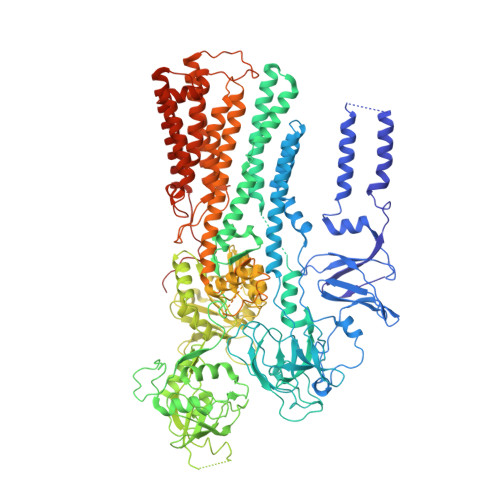
6XMU

Structure of P5A-ATPase Spf1, endogenous substrate-bound


Experimental Data Snapshot

  • Method: ELECTRON MICROSCOPY
  • Resolution: 3.30 Å
  • Aggregation State: PARTICLE 
  • Reconstruction Method: SINGLE PARTICLE 

wwPDB Validation   3D Report Full Report


This is version 1.2 of the entry. See complete history


Literature

The endoplasmic reticulum P5A-ATPase is a transmembrane helix dislocase.

McKenna, M.J.Sim, S.I.Ordureau, A.Wei, L.Harper, J.W.Shao, S.Park, E.

(2020) Science 369

  • DOI: https://doi.org/10.1126/science.abc5809
  • Primary Citation of Related Structures:  
    6XMP, 6XMQ, 6XMS, 6XMT, 6XMU

  • PubMed Abstract: 

    Organelle identity depends on protein composition. How mistargeted proteins are selectively recognized and removed from organelles is incompletely understood. Here, we found that the orphan P5A-adenosine triphosphatase (ATPase) transporter ATP13A1 (Spf1 in yeast) directly interacted with the transmembrane segment (TM) of mitochondrial tail-anchored proteins. P5A-ATPase activity mediated the extraction of mistargeted proteins from the endoplasmic reticulum (ER). Cryo-electron microscopy structures of Saccharomyces cerevisiae Spf1 revealed a large, membrane-accessible substrate-binding pocket that alternately faced the ER lumen and cytosol and an endogenous substrate resembling an α-helical TM. Our results indicate that the P5A-ATPase could dislocate misinserted hydrophobic helices flanked by short basic segments from the ER. TM dislocation by the P5A-ATPase establishes an additional class of P-type ATPase substrates and may correct mistakes in protein targeting or topogenesis.


  • Organizational Affiliation

    Department of Cell Biology, Blavatnik Institute, Harvard Medical School, Boston, MA 02115, USA.


Macromolecules
Find similar proteins by:  (by identity cutoff)  |  3D Structure
Entity ID: 1
MoleculeChains Sequence LengthOrganismDetailsImage
P5A-type ATPase1,239Saccharomyces cerevisiae S288CMutation(s): 0 
EC: 7.2.2
Membrane Entity: Yes 
UniProt
Find proteins for P39986 (Saccharomyces cerevisiae (strain ATCC 204508 / S288c))
Explore P39986 
Go to UniProtKB:  P39986
Entity Groups  
Sequence Clusters30% Identity50% Identity70% Identity90% Identity95% Identity100% Identity
UniProt GroupP39986
Sequence Annotations
Expand
  • Reference Sequence

Find similar proteins by:  Sequence   |   3D Structure  

Entity ID: 2
MoleculeChains Sequence LengthOrganismDetailsImage
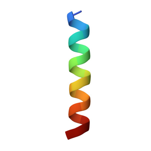
Putative endogenous substrate transmembrane helix20Saccharomyces cerevisiae S288CMutation(s): 0 
Sequence Annotations
Expand
  • Reference Sequence
Small Molecules
Experimental Data & Validation

Experimental Data

  • Method: ELECTRON MICROSCOPY
  • Resolution: 3.30 Å
  • Aggregation State: PARTICLE 
  • Reconstruction Method: SINGLE PARTICLE 
EM Software:
TaskSoftware PackageVersion
RECONSTRUCTIONcryoSPARC2.12.4

Structure Validation

View Full Validation Report


Entry History & Funding Information

Deposition Data


Funding OrganizationLocationGrant Number
Other privateUnited States--

Revision History  (Full details and data files)

  • Version 1.0: 2020-09-23
    Type: Initial release
  • Version 1.1: 2020-10-07
    Changes: Database references
  • Version 1.2: 2024-03-06
    Changes: Data collection, Database references