Structure-Aided Development of Small-Molecule Inhibitors of ENPP1, the Extracellular Phosphodiesterase of the Immunotransmitter cGAMP.
Carozza, J.A., Brown, J.A., Bohnert, V., Fernandez, D., AlSaif, Y., Mardjuki, R.E., Smith, M., Li, L.(2020) Cell Chem Biol 27: 1347-1358.e5
- PubMed: 32726585 Search on PubMedSearch on PubMed Central
- DOI: https://doi.org/10.1016/j.chembiol.2020.07.007
- Primary Citation Related Structures:
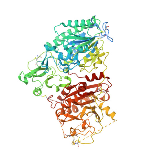
6XKD - PubMed Abstract:
Cancer cells initiate an innate immune response by synthesizing and exporting the small-molecule immunotransmitter cGAMP, which activates the anti-cancer Stimulator of Interferon Genes (STING) pathway in the host. An extracellular enzyme, ectonucleotide pyrophosphatase phosphodiesterase 1 (ENPP1), hydrolyzes cGAMP and negatively regulates this anti-cancer immune response. Small-molecule ENPP1 inhibitors are much needed as tools to study the basic biology of extracellular cGAMP and as investigational cancer immunotherapy drugs. Here, we surveyed structure-activity relationships around a series of cell-impermeable and thus extracellular-targeting phosphonate inhibitors of ENPP1. In addition, we solved the crystal structure of an exemplary phosphonate inhibitor to elucidate the interactions that drive potency. This study yielded several best-in-class inhibitors with K i < 2 nM and excellent physicochemical and pharmacokinetic properties. Finally, we demonstrate that an ENPP1 inhibitor delays tumor growth in a breast cancer mouse model. Together, we have developed ENPP1 inhibitors that are excellent tool compounds and potential therapeutics.
- Department of Chemistry, Stanford University, Stanford, CA 93405, USA; Stanford ChEM-H, Stanford University, Stanford, CA 93405, USA.
Organizational Affiliation: