6XJQ

Crystal structure of a self-alkylating ribozyme - alkylated form with biotinylated epoxide substrate


Experimental Data Snapshot

  • Method: X-RAY DIFFRACTION
  • Resolution: 1.71 Å
  • R-Value Free: 0.213 
  • R-Value Work: 0.189 
  • R-Value Observed: 0.189 

wwPDB Validation   3D Report Full Report


Ligand Structure Quality Assessment 


This is version 1.3 of the entry. See complete history


Literature

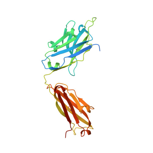
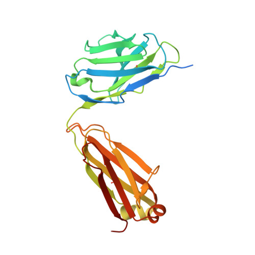
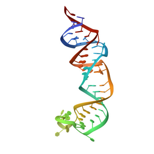
Structural basis for substrate binding and catalysis by a self-alkylating ribozyme.

Krochmal, D.Shao, Y.Li, N.S.DasGupta, S.Shelke, S.A.Koirala, D.Piccirilli, J.A.

(2022) Nat Chem Biol 18: 376-384

  • DOI: https://doi.org/10.1038/s41589-021-00950-z
  • Primary Citation of Related Structures:  
    6XJQ, 6XJW, 6XJY, 6XJZ

  • PubMed Abstract: 

    Ribozymes that react with small-molecule probes have important applications in transcriptomics and chemical biology, such as RNA labeling and imaging. Understanding the structural basis for these RNA-modifying reactions will enable the development of better tools for studying RNA. Nevertheless, high-resolution structures and underlying catalytic mechanisms for members of this ribozyme class remain elusive. Here, we focus on a self-alkylating ribozyme that catalyzes nitrogen-carbon bond formation between a specific guanine and a 2,3-disubstituted epoxide substrate and report the crystal structures of a self-alkylating ribozyme, including both alkylated and apo forms, at 1.71-Å and 2.49-Å resolution, respectively. The ribozyme assumes an elongated hairpin-like architecture preorganized to accommodate the epoxide substrate in a hook-shaped conformation. Observed reactivity of substrate analogs together with an inverse, log-linear pH dependence of the reaction rate suggests a requirement for epoxide protonation, possibly assisted by the ether oxygens within the substrate.


  • Organizational Affiliation

    Department of Biochemistry and Molecular Biology, The University of Chicago, Chicago, IL, USA.


Macromolecules

Find similar proteins by:  (by identity cutoff)  |  3D Structure
Entity ID: 2
MoleculeChains Sequence LengthOrganismDetailsImage
Fab HAVx Heavy ChainC,
E [auth H]
258Homo sapiensMutation(s): 0 
Entity Groups  
Sequence Clusters30% Identity50% Identity70% Identity90% Identity95% Identity100% Identity
Sequence Annotations
Expand
  • Reference Sequence
Find similar proteins by:  (by identity cutoff)  |  3D Structure
Entity ID: 3
MoleculeChains Sequence LengthOrganismDetailsImage
Fab HAVx Light ChainD,
F [auth L]
238Homo sapiensMutation(s): 0 
Entity Groups  
Sequence Clusters30% Identity50% Identity70% Identity90% Identity95% Identity100% Identity
Sequence Annotations
Expand
  • Reference Sequence
Find similar nucleic acids by:  (by identity cutoff)  |  3D Structure
Entity ID: 1
MoleculeChains LengthOrganismImage
Self-alkylating ribozyme (58-MER)
A, B
58Aeropyrum pernix
Sequence Annotations
Expand
  • Reference Sequence
Small Molecules
Ligands 1 Unique
IDChains Name / Formula / InChI Key2D Diagram3D Interactions
V4J (Subject of Investigation/LOI)
Query on V4J

Download Ideal Coordinates CCD File 
G [auth A],
H [auth B]
2-{[(4R)-4-hydroxyhexyl]oxy}ethyl 5-[(3aS,4S,6aR)-2-oxohexahydro-1H-thieno[3,4-d]imidazol-4-yl]pentanoate
C18 H32 N2 O5 S
XPLYDBRQTYFSEO-AESZEHBQSA-N
Experimental Data & Validation

Experimental Data

  • Method: X-RAY DIFFRACTION
  • Resolution: 1.71 Å
  • R-Value Free: 0.213 
  • R-Value Work: 0.189 
  • R-Value Observed: 0.189 
  • Space Group: P 1 21 1
Unit Cell:
Length ( Å )Angle ( ˚ )
a = 83.962α = 90
b = 99.832β = 97.2
c = 90.636γ = 90
Software Package:
Software NamePurpose
PHENIXrefinement
XDSdata scaling
PDB_EXTRACTdata extraction
XDSdata reduction
PHENIXphasing

Structure Validation

View Full Validation Report


Ligand Structure Quality Assessment 


Entry History & Funding Information

Deposition Data


Funding OrganizationLocationGrant Number
National Institutes of Health/National Human Genome Research Institute (NIH/NHGRI)United StatesR01AI081987
National Institutes of Health/National Human Genome Research Institute (NIH/NHGRI)United StatesR01GM102489

Revision History  (Full details and data files)

  • Version 1.0: 2022-01-19
    Type: Initial release
  • Version 1.1: 2022-02-09
    Changes: Database references
  • Version 1.2: 2022-04-13
    Changes: Database references
  • Version 1.3: 2023-10-18
    Changes: Data collection, Refinement description