6XIT

Cryo-EM structure of the G protein-gated inward rectifier K+ channel GIRK2 (Kir3.2) in complex with PIP2


Experimental Data Snapshot

  • Method: ELECTRON MICROSCOPY
  • Resolution: 3.30 Å
  • Aggregation State: PARTICLE 
  • Reconstruction Method: SINGLE PARTICLE 

wwPDB Validation   3D Report Full Report


This is version 1.0 of the entry. See complete history


Literature

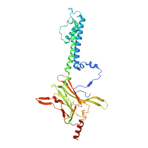
Cryo-EM analysis of PIP 2 regulation in mammalian GIRK channels.

Niu, Y.Tao, X.Touhara, K.K.MacKinnon, R.

(2020) Elife 9

  • DOI: https://doi.org/10.7554/eLife.60552
  • Primary Citation of Related Structures:  
    6XIS, 6XIT

  • PubMed Abstract: 

    G-protein-gated inward rectifier potassium (GIRK) channels are regulated by G proteins and PIP 2 . Here, using cryo-EM single particle analysis we describe the equilibrium ensemble of structures of neuronal GIRK2 as a function of the C8-PIP 2 concentration. We find that PIP 2 shifts the equilibrium between two distinguishable structures of neuronal GIRK (GIRK2), extended and docked, towards the docked form. In the docked form the cytoplasmic domain, to which G βγ binds, becomes accessible to the cytoplasmic membrane surface where G βγ resides. Furthermore, PIP 2 binding reshapes the G βγ binding surface on the cytoplasmic domain, preparing it to receive G βγ . We find that cardiac GIRK (GIRK1/4) can also exist in both extended and docked conformations. These findings lead us to conclude that PIP 2 influences GIRK channels in a structurally similar manner to Kir2.2 channels. In Kir2.2 channels, the PIP 2 -induced conformational changes open the pore. In GIRK channels, they prepare the channel for activation by G βγ .


  • Organizational Affiliation

    Laboratory of Molecular Neurobiology and Biophysics, The Rockefeller University, Howard Hughes Medical Institute, New York, United States.


Macromolecules
Find similar proteins by:  (by identity cutoff)  |  3D Structure
Entity ID: 1
MoleculeChains Sequence LengthOrganismDetailsImage
G protein-activated inward rectifier potassium channel 2
A, B, C, D
340Mus musculusMutation(s): 0 
Gene Names: Kcnj6
Membrane Entity: Yes 
UniProt & NIH Common Fund Data Resources
Find proteins for P48542 (Mus musculus)
Explore P48542 
Go to UniProtKB:  P48542
IMPC:  MGI:104781
Entity Groups  
Sequence Clusters30% Identity50% Identity70% Identity90% Identity95% Identity100% Identity
UniProt GroupP48542
Sequence Annotations
Expand
  • Reference Sequence
Experimental Data & Validation

Experimental Data

  • Method: ELECTRON MICROSCOPY
  • Resolution: 3.30 Å
  • Aggregation State: PARTICLE 
  • Reconstruction Method: SINGLE PARTICLE 
EM Software:
TaskSoftware PackageVersion
RECONSTRUCTIONRELION3.1
MODEL REFINEMENTPHENIX1.15.2

Structure Validation

View Full Validation Report


Entry History & Funding Information

Deposition Data


Funding OrganizationLocationGrant Number
National Institutes of Health/National Institute of General Medical Sciences (NIH/NIGMS)United StatesGM43949

Revision History  (Full details and data files)

  • Version 1.0: 2020-10-07
    Type: Initial release