6XHK

Crystal structure of S. aureus TarJ in complex with NADPH


Experimental Data Snapshot

  • Method: X-RAY DIFFRACTION
  • Resolution: 3.00 Å
  • R-Value Free: 0.262 
  • R-Value Work: 0.205 
  • R-Value Observed: 0.208 

wwPDB Validation   3D Report Full Report


Ligand Structure Quality Assessment 


This is version 1.2 of the entry. See complete history


Literature

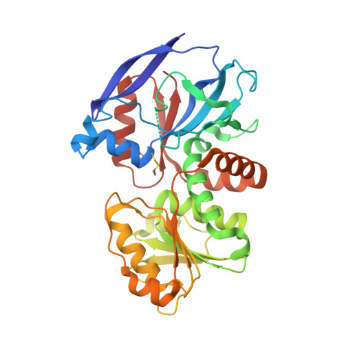
Crystallographic analysis of TarI and TarJ, a cytidylyltransferase and reductase pair for CDP-ribitol synthesis in Staphylococcus aureus wall teichoic acid biogenesis.

Li, F.K.K.Gale, R.T.Petrotchenko, E.V.Borchers, C.H.Brown, E.D.Strynadka, N.C.J.

(2021) J Struct Biol 213: 107733-107733

  • DOI: https://doi.org/10.1016/j.jsb.2021.107733
  • Primary Citation of Related Structures:  
    6XH9, 6XHK, 6XHP, 6XHQ, 6XHR, 6XHS, 6XHT

  • PubMed Abstract: 

    The cell wall of many pathogenic Gram-positive bacteria contains ribitol-phosphate wall teichoic acid (WTA), a polymer that is linked to virulence and regulation of essential physiological processes including cell division. CDP-ribitol, the activated precursor for ribitol-phosphate polymerization, is synthesized by a cytidylyltransferase and reductase pair known as TarI and TarJ, respectively. In this study, we present crystal structures of Staphylococcus aureus TarI and TarJ in their apo forms and in complex with substrates and products. The TarI structures illustrate the mechanism of CDP-ribitol synthesis from CTP and ribitol-phosphate and reveal structural changes required for substrate binding and catalysis. Insights into the upstream step of ribulose-phosphate reduction to ribitol-phosphate is provided by the structures of TarJ. Furthermore, we propose a general topology of the enzymes in a heterotetrameric form built using restraints from crosslinking mass spectrometry analysis. Together, our data present molecular details of CDP-ribitol production that may aid in the design of inhibitors against WTA biosynthesis.


  • Organizational Affiliation

    Department of Biochemistry and Molecular Biology and Centre for Blood Research, The University of British Columbia, Vancouver, British Columbia V6T 1Z3, Canada.


Macromolecules
Find similar proteins by:  (by identity cutoff)  |  3D Structure
Entity ID: 1
MoleculeChains Sequence LengthOrganismDetailsImage
Ribulose-5-phosphate reductase 1341Staphylococcus aureus subsp. aureus NCTC 8325Mutation(s): 0 
Gene Names: tarJSAOUHSC_00226
EC: 1.1.1.405
UniProt
Find proteins for Q2G1B9 (Staphylococcus aureus (strain NCTC 8325 / PS 47))
Explore Q2G1B9 
Go to UniProtKB:  Q2G1B9
Entity Groups  
Sequence Clusters30% Identity50% Identity70% Identity90% Identity95% Identity100% Identity
UniProt GroupQ2G1B9
Sequence Annotations
Expand
  • Reference Sequence
Small Molecules
Ligands 1 Unique
IDChains Name / Formula / InChI Key2D Diagram3D Interactions
NDP
Query on NDP

Download Ideal Coordinates CCD File 
B [auth A]NADPH DIHYDRO-NICOTINAMIDE-ADENINE-DINUCLEOTIDE PHOSPHATE
C21 H30 N7 O17 P3
ACFIXJIJDZMPPO-NNYOXOHSSA-N
Modified Residues  1 Unique
IDChains TypeFormula2D DiagramParent
CME
Query on CME
A
L-PEPTIDE LINKINGC5 H11 N O3 S2CYS
Experimental Data & Validation

Experimental Data

  • Method: X-RAY DIFFRACTION
  • Resolution: 3.00 Å
  • R-Value Free: 0.262 
  • R-Value Work: 0.205 
  • R-Value Observed: 0.208 
  • Space Group: P 62 2 2
Unit Cell:
Length ( Å )Angle ( ˚ )
a = 99.237α = 90
b = 99.237β = 90
c = 268.757γ = 120
Software Package:
Software NamePurpose
PHENIXrefinement
PDB_EXTRACTdata extraction
XDSdata reduction
Aimlessdata scaling
PHASERphasing

Structure Validation

View Full Validation Report


Ligand Structure Quality Assessment 


Entry History & Funding Information

Deposition Data


Funding OrganizationLocationGrant Number
Canadian Institutes of Health Research (CIHR)Canada--
Howard Hughes Medical Institute (HHMI)Canada--
Natural Sciences and Engineering Research Council (NSERC, Canada)Canada--

Revision History  (Full details and data files)

  • Version 1.0: 2021-04-21
    Type: Initial release
  • Version 1.1: 2021-05-05
    Changes: Database references
  • Version 1.2: 2023-10-18
    Changes: Data collection, Database references, Refinement description