Going Platinum to the Tune of a Remarkable Guanine Quadruplex Binder: Solution- and Solid-State Investigations.
Miron, C.E., van Staalduinen, L., Rangaswamy, A.M., Chen, M., Liang, Y., Jia, Z., Mergny, J.L., Petitjean, A.(2021) Angew Chem Int Ed Engl 60: 2500-2507
- PubMed: 33090592 Search on PubMed
- DOI: https://doi.org/10.1002/anie.202012520
- Primary Citation Related Structures:
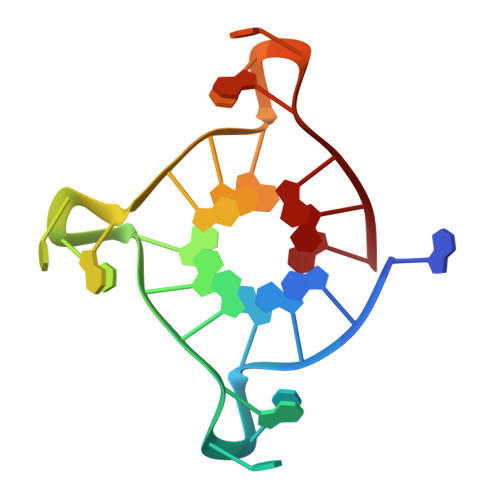
6XCL - PubMed Abstract:
Guanine quadruplex recognition has gained increasing attention, inspired by the growing awareness of the key roles played by these non-canonical nucleic acid architectures in cellular regulatory processes. We report here the solution and solid-state studies of a novel planar platinum(II) complex that is easily assembled from a simple ligand, and exhibits notable binding affinity for guanine quadruplex structures, while maintaining good selectivity for guanine quadruplex over duplex structures. A crystal structure of this ligand complexed with a telomeric quadruplex confirms double end-capping, with dimerization at the 5' interface.
- Department of Chemistry, Queen's University, 90 Bader Lane, Kingston, ON, K7L 3N6, Canada.
Organizational Affiliation: