6WS2

Crystal structure of KRAS-K104Q mutant, GDP-bound


Experimental Data Snapshot

  • Method: X-RAY DIFFRACTION
  • Resolution: 1.59 Å
  • R-Value Free: 0.195 
  • R-Value Work: 0.182 
  • R-Value Observed: 0.182 

wwPDB Validation   3D Report Full Report


Ligand Structure Quality Assessment 


This is version 1.2 of the entry. See complete history


Literature

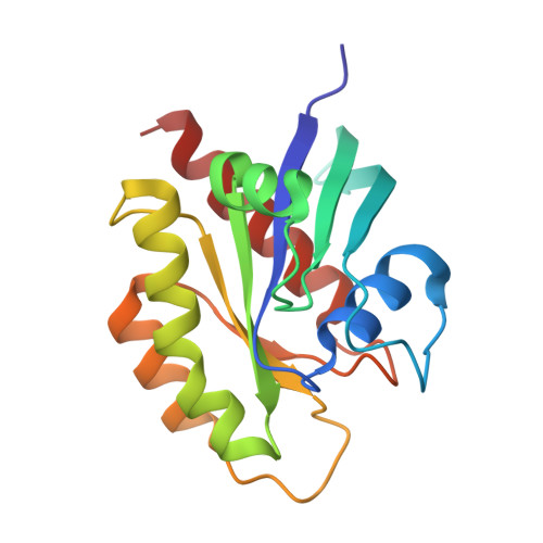
Allosteric regulation of switch-II domain controls KRAS oncogenicity.

Yang, M.H.Tran, T.H.Hunt, B.Agnor, R.Johnson, C.W.Shui, B.Waybright, T.J.Nowak, J.A.Stephen, A.G.Simanshu, D.K.Haigis, K.M.

(2023) Cancer Res 

  • DOI: https://doi.org/10.1158/0008-5472.CAN-22-3210
  • Primary Citation of Related Structures:  
    6WS2, 6WS4, 8STM, 8STN

  • PubMed Abstract: 

    RAS proteins are GTPases that regulate a wide range of cellular processes. RAS activity is dependent on its nucleotide-binding status, which is modulated by guanine nucleotide exchange factors (GEF) and GTPase-activating proteins (GAP). KRAS can be acetylated at lysine 104 (K104), and an acetylation-mimetic mutation of K104 to glutamine (K104Q) attenuates the in vitro-transforming capacity of oncogenic KRAS by interrupting GEF-induced nucleotide exchange. To assess the effect of this mutation in vivo, we used CRISPR-Cas9 to generate mouse models carrying the K104Q point mutation in wild-type and conditional KrasLSL-G12D alleles. Homozygous animals for K104Q were viable, fertile, and arose at the expected Mendelian frequency, indicating that K104Q is not a complete loss-of-function mutation. Consistent with our previous findings from in vitro studies, however, the oncogenic activity of KRASG12D was significantly attenuated by mutation at K104. Biochemical and structural analysis indicated that the G12D and K104Q mutations cooperate to suppress GEF-mediated nucleotide exchange, explaining the preferential effect of K104Q on oncogenic KRAS. Furthermore, K104 functioned in an allosteric network with M72, R73, and G75 on the α2 helix of the switch-II region. Intriguingly, point mutation of glycine 75 to alanine (G75A) also showed a strong negative regulatory effect on KRASG12D. These data demonstrate that lysine at position 104 is critical for the full oncogenic activity of mutant KRAS and suggest that modulating the sites in its allosteric network may provide a unique therapeutic approach in cancers expressing mutant KRAS.


  • Organizational Affiliation

    Department of Cancer Biology, Dana-Farber Cancer Institute, Boston, Massachusetts.


Macromolecules
Find similar proteins by:  (by identity cutoff)  |  3D Structure
Entity ID: 1
MoleculeChains Sequence LengthOrganismDetailsImage
GTPase KRas
A, B, C, D
170Homo sapiensMutation(s): 1 
Gene Names: KRASKRAS2RASK2
UniProt & NIH Common Fund Data Resources
Find proteins for P01116 (Homo sapiens)
Explore P01116 
Go to UniProtKB:  P01116
PHAROS:  P01116
GTEx:  ENSG00000133703 
Entity Groups  
Sequence Clusters30% Identity50% Identity70% Identity90% Identity95% Identity100% Identity
UniProt GroupP01116
Sequence Annotations
Expand
  • Reference Sequence
Small Molecules
Ligands 4 Unique
IDChains Name / Formula / InChI Key2D Diagram3D Interactions
GDP (Subject of Investigation/LOI)
Query on GDP

Download Ideal Coordinates CCD File 
E [auth A],
K [auth B],
P [auth C],
S [auth D]
GUANOSINE-5'-DIPHOSPHATE
C10 H15 N5 O11 P2
QGWNDRXFNXRZMB-UUOKFMHZSA-N
SO4
Query on SO4

Download Ideal Coordinates CCD File 
G [auth A]
H [auth A]
I [auth A]
J [auth A]
M [auth B]
G [auth A],
H [auth A],
I [auth A],
J [auth A],
M [auth B],
N [auth B],
R [auth C]
SULFATE ION
O4 S
QAOWNCQODCNURD-UHFFFAOYSA-L
CL
Query on CL

Download Ideal Coordinates CCD File 
O [auth B]CHLORIDE ION
Cl
VEXZGXHMUGYJMC-UHFFFAOYSA-M
MG (Subject of Investigation/LOI)
Query on MG

Download Ideal Coordinates CCD File 
F [auth A],
L [auth B],
Q [auth C],
T [auth D]
MAGNESIUM ION
Mg
JLVVSXFLKOJNIY-UHFFFAOYSA-N
Experimental Data & Validation

Experimental Data

  • Method: X-RAY DIFFRACTION
  • Resolution: 1.59 Å
  • R-Value Free: 0.195 
  • R-Value Work: 0.182 
  • R-Value Observed: 0.182 
  • Space Group: P 3
Unit Cell:
Length ( Å )Angle ( ˚ )
a = 84.56α = 90
b = 84.56β = 90
c = 88.36γ = 120
Software Package:
Software NamePurpose
XDSdata reduction
XSCALEdata scaling
REFMACrefinement
PDB_EXTRACTdata extraction
PHASERphasing

Structure Validation

View Full Validation Report


Ligand Structure Quality Assessment 


Entry History & Funding Information

Deposition Data


Funding OrganizationLocationGrant Number
National Institutes of Health/National Cancer Institute (NIH/NCI)United StatesHHSN261200800001E

Revision History  (Full details and data files)

  • Version 1.0: 2021-11-10
    Type: Initial release
  • Version 1.1: 2023-08-23
    Changes: Data collection, Database references, Refinement description
  • Version 1.2: 2023-10-18
    Changes: Refinement description