


Crystal structure of Aspartyl-tRNA ligase from Elizabethkingia sp.
Abendroth, J., Lorimer, D.D., Horanyi, P.S., Edwards, T.E.To be published.
Experimental Data Snapshot
Starting Model: experimental
View more details
wwPDB Validation 3D Report Full Report

Entity ID: 1 | |||||
|---|---|---|---|---|---|
| Molecule | Chains | Sequence Length | Organism | Details | Image |
| Aspartate--tRNA ligase | 592 | Elizabethkingia anophelis | Mutation(s): 0 Gene Names: aspS, AYC66_05975, BAY09_04240, BAY10_00500 EC: 6.1.1.12 | 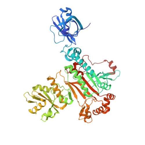 | |
UniProt | |||||
Find proteins for A0A1T3DDI2 (Elizabethkingia anophelis) Explore A0A1T3DDI2 Go to UniProtKB: A0A1T3DDI2 | |||||
Entity Groups | |||||
| Sequence Clusters | 30% Identity50% Identity70% Identity90% Identity95% Identity100% Identity | ||||
| UniProt Group | A0A1T3DDI2 | ||||
Sequence AnnotationsExpand | |||||
Reference Sequence | |||||
| Ligands 1 Unique | |||||
|---|---|---|---|---|---|
| ID | Chains | Name / Formula / InChI Key | 2D Diagram | 3D Interactions | |
| SO4 Download:Ideal Coordinates CCD File | D [auth A], E [auth B] | SULFATE ION O4 S QAOWNCQODCNURD-UHFFFAOYSA-L |  | ||
| Length ( Å ) | Angle ( ˚ ) |
|---|---|
| a = 234.37 | α = 90 |
| b = 106.57 | β = 100.91 |
| c = 93.41 | γ = 90 |
| Software Name | Purpose |
|---|---|
| XDS | data reduction |
| XSCALE | data scaling |
| PHENIX | refinement |
| PDB_EXTRACT | data extraction |
| MoRDa | phasing |
| ARP/wARP | model building |
| Coot | model building |


